 Explore
 Validate
 Learn
Learn Western blot
 ELISA
ELISAAntibody data
- Antibody Data
- Antigen structure
- References [0]
- Comments [0]
- Validations
- Western blot [2]
- Immunohistochemistry [8]
Submit
Validation data
Reference
Comment
Report error
- Product number
- ABIN2506482 - Provider product page

- Provider
- antibodies-online
- Product name
- anti-TAR DNA Binding Protein (TARDBP) antibody
- Antibody type
- Polyclonal
- Antigen
- The immunogen for anti-TDP43 antibody: antibody was raised against a 18 amino acid peptide from near the center of human TDP43.
- Description
- Affinity chromatography purified via peptide column
- Reactivity
- Human, Mouse, Rat
- Host
- Rabbit
- Vial size
- 0.1 mg
- Storage
- TDP43 antibody can be stored at 4°C, stable for one year. As with all antibodies care should be taken to avoid repeated freeze thaw cycles. Antibodies should not be exposed to prolonged high temperatures.
- Handling
- As with all antibodies care should be taken to avoid repeated freeze thaw cycles. Antibodies should not be exposed to prolonged high temperatures.
No comments: Submit comment
Supportive validation
- Submitted by
- antibodies-online (provider)
- Main image
- Experimental details
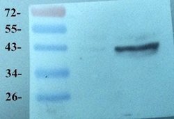
- WB analysis of Rat brain tissue using Melatonin Receptor (dilution of primary antibody - 2 ug/ml)

- Submitted by
- antibodies-online (provider)
- Main image

- Experimental details
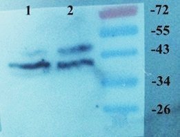
- Western blot analysis of Rat brain (lane 1), Mouse brain (lane 2) using Melatonin Receptor antibody (Dilution at 2 ug/ml)
Supportive validation
- Submitted by
- antibodies-online (provider)
- Main image
- Experimental details
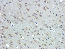
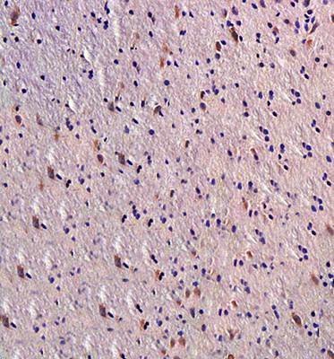
- IHC-P image of rat brain tissue using anti-Melatonin receptor 1A (dilution of primary antibody at 1:500)
- Submitted by
- antibodies-online (provider)
- Main image

- Experimental details
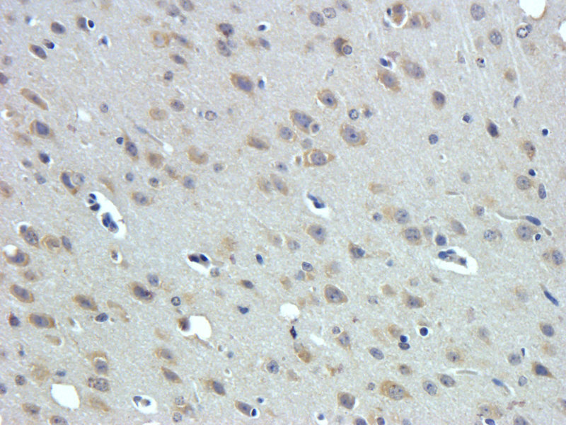
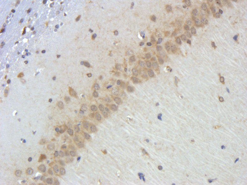
- IHC-P staining of rat brain hippocampus layer tissue using anti-Melatonin receptor 1A (dilution at 1:500)
- Submitted by
- antibodies-online (provider)
- Main image

- Experimental details
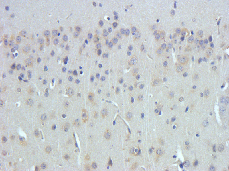
- Immunohistrochemical analysis of formalin fixed and paraffin embedded rat brain tissue(Snr) using ant Melatonin Receptor 1A (primary antibody at 1:300)
- Submitted by
- antibodies-online (provider)
- Main image

- Experimental details
- Immunohistochemical staining of rat brain tissue using Melatonin receptor 1A antibody (dilution of primary antibody - 1:500)
- Submitted by
- antibodies-online (provider)
- Main image

- Experimental details
- IHC-P of mouse hippocampus tissue (orb11085 at 1:300)

- Submitted by
- antibodies-online (provider)
- Main image

- Experimental details
- IHC-P of mouse hippocampus tissue using Melatonin Receptor 1A antibody (orb11085 at 1:300)

- Submitted by
- antibodies-online (provider)
- Main image

- Experimental details
- Immunohistochemical staining of mouse brain tissue using Melatonin receptor 1A antibody (dilution of primary antibody - 1:300)

- Submitted by
- antibodies-online (provider)
- Main image

- Experimental details
- Immunohistochemical staining of paraffin embedded mouse hippocampus tissue using Melatonin Receptor 1A antibody (dilution at 1:500)
