Explore
 Validate
 Learn
Learn Western blot
Antibody data
- Antibody Data
- Antigen structure
- References [0]
- Comments [0]
- Validations
- Western blot [3]
- Immunocytochemistry [4]
Submit
Validation data
Reference
Comment
Report error
- Product number
- PA5-17299 - Provider product page

- Provider
- Invitrogen Antibodies
- Product name
- PRMT1 Polyclonal Antibody
- Antibody type
- Polyclonal
- Antigen
- Synthetic peptide
- Description
- It is not recommended to aliquot this antibody.
- Concentration
- 5 µg/mL
No comments: Submit comment
Supportive validation
- Submitted by
- Invitrogen Antibodies (provider)
- Main image
- Experimental details
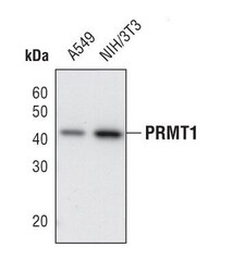
- Western blot analysis of PRMT1 in cell lysates from A549 and NIH/3T3 cells using PRMT1 polyclonal antibody (Product # PA5-17299).
- Submitted by
- Invitrogen Antibodies (provider)
- Main image
- Experimental details
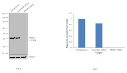
- Knockdown of PRMT1 was achieved by transfecting HCT 116 cells with PRMT1 specific siRNAs (Silencer® select Product # s6918; s6917). Western blot analysis (Fig. a) was performed using modified whole cell extracts (1% SDS) from the PRMT1 knockdown cells (lane 3), non-specific scrambled siRNA transfected cells (lane 2) and untransfected cells (lane 1). The blots were probed with PRMT1 Polyclonal Antibody (Product # PA5-17299, 1:1000 dilution) and Goat anti-Rabbit IgG (H+L) Superclonal™ Secondary Antibody, HRP conjugate (Product # A27036, 0.25 µg/mL, 1:4000 dilution). Densitometric analysis of this western blot is shown in histogram (Fig. b). Decrease in signal upon siRNA mediated knock down confirms that antibody is specific to PRMT1.

- Submitted by
- Invitrogen Antibodies (provider)
- Main image
- Experimental details
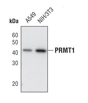
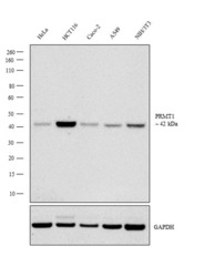
- Western blot analysis was performed on modified whole cell extracts (15 SDS) (30 µg lysate) of HeLa (Lane 1), HCT116 (Lane 2), Caco-2 (Lane 3), A549 (Lane 4), and NIH/3T3 (Lane 5). The blot was probed with Anti-PRMT1 Polyclonal Antibody (Product # PA5-17299, 1:1000 dilution) and detected by chemiluminescence using Goat anti-Rabbit IgG (H+L) Superclonal™ Secondary Antibody, HRP conjugate (Product # A27036, 0.25 µg/mL, 1:4000 dilution). A 42 kDa band corresponding to PRMT1 was detected in all human and mouse cell line(s) tested.

Supportive validation
- Submitted by
- Invitrogen Antibodies (provider)
- Main image

- Experimental details
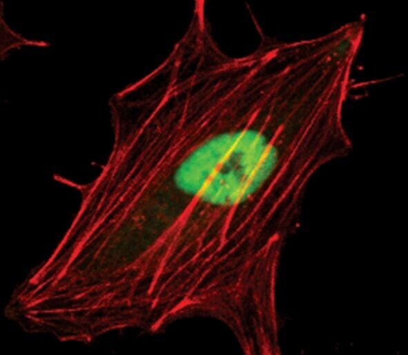
- Immunofluorescent analysis of PRMT1 in either untreated HeLa using a PRMT1 polyclonal antibody (Product # PA5-17299) (green). Actin filaments are labeled with a fluorescent red phalloidin.
- Submitted by
- Invitrogen Antibodies (provider)
- Main image
- Experimental details
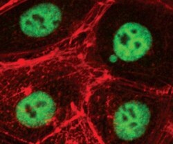
- Immunofluorescent analysis of PRMT1 in either untreated HCT-116 using a PRMT1 polyclonal antibody (Product # PA5-17299) (green). Actin filaments are labeled with a fluorescent red phalloidin.

- Submitted by
- Invitrogen Antibodies (provider)
- Main image

- Experimental details
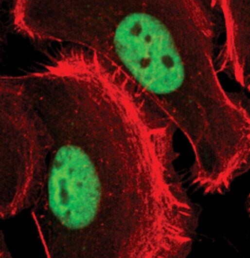
- Immunofluorescent analysis of PRMT1 in either untreated SK-OV-3 using a PRMT1 polyclonal antibody (Product # PA5-17299) (green). Actin filaments are labeled with a fluorescent red phalloidin.
- Submitted by
- Invitrogen Antibodies (provider)
- Main image
- Experimental details
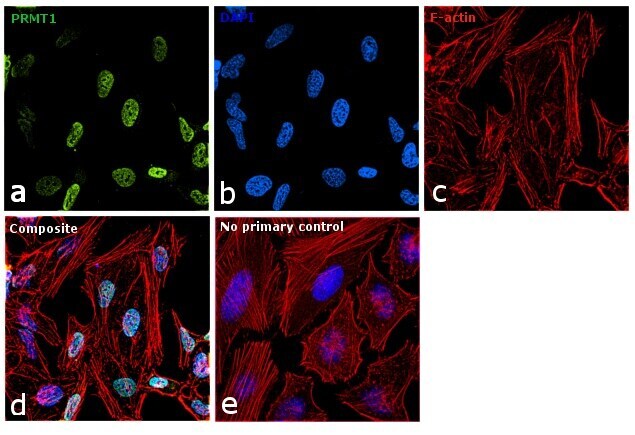
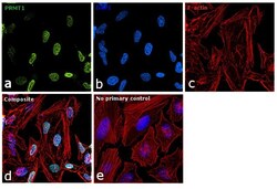
- Immunofluorescence analysis of PRMT1 was performed using 70% confluent log phase HeLa cells. The cells were fixed with 4% paraformaldehyde for 10 minutes, permeabilized with 0.1% Triton™ X-100 for 15 minutes, and blocked with 1% BSA for 1 hour at room temperature. The cells were labeled with PRMT1 Polyclonal Antibody (Product # PA5-17299) at 1:200 dilution in 0.1% BSA, incubated at 4 degree Celsius overnight and then labeled with Goat anti-Rabbit IgG (H+L) Superclonal™ Secondary Antibody, Alexa Fluor® 488 conjugate (Product # A27034) at a dilution of 1:2000 for 45 minutes at room temperature (Panel a: green). Nuclei (Panel b: blue) were stained with ProLong™ Diamond Antifade Mountant with DAPI (Product # P36962). F-actin (Panel c: red) was stained with Rhodamine Phalloidin (Product # R415). Panel d represents the merged image showing Nuclear localization. Panel e represents control cells with no primary antibody to assess background. The images were captured at 60X magnification.