 Explore
 Validate
 Learn
LearnPA1-32476
antibody from Invitrogen Antibodies
Targeting: GFRA1
GDNFR, GDNFRA, GFR-ALPHA-1, RET1L, RETL1, TRNR1
 Western blot
 Immunocytochemistry
Antibody data
- Antibody Data
- Antigen structure
- References [1]
- Comments [0]
- Validations
- Immunocytochemistry [1]
Submit
Validation data
Reference
Comment
Report error
- Product number
- PA1-32476 - Provider product page

- Provider
- Invitrogen Antibodies
- Product name
- GFR alpha-1 Polyclonal Antibody
- Antibody type
- Polyclonal
- Antigen
- Synthetic peptide
- Description
- Super Bright 436 can be excited with the violet laser line (405 nm) and emits at 436 nm. We recommend using a 450/50 bandpass filter, or equivalent. Please make sure that your instrument is capable of detecting this fluorochrome.
- Reactivity
- Human, Mouse, Rat
- Host
- Rabbit
- Isotype
- IgG
- Vial size
- 100 µg
- Concentration
- 1 mg/mL
- Storage
- Store at 4°C short term. For long term storage, store at -20°C, avoiding freeze/thaw cycles.
Submitted references Cytotoxicity of nonylphenol on spermatogonial stem cells via phosphatidylinositol-3-kinase/protein kinase B/mammalian target of rapamycin pathway.
Lei JH, Yan W, Luo CH, Guo YM, Zhang YY, Wang XH, Su XJ
World journal of stem cells 2020 Jun 26;12(6):500-513
World journal of stem cells 2020 Jun 26;12(6):500-513
No comments: Submit comment
Supportive validation
- Submitted by
- Invitrogen Antibodies (provider)
- Main image
- Experimental details
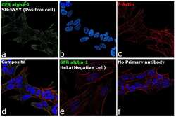
- Immunofluorescence analysis of GFR alpha-1 was performed using SH-SY5Y and HeLa cells. The cells were fixed with 4% paraformaldehyde for 10 minutes, permeabilized with 0.1% Triton™ X-100 for 15 minutes, and blocked with 2% BSA for 1 hour at room temperature. The cells were labeled with GFR alpha-1 Polyclonal Antibody (Product # PA1-32476) at 1:100 dilution in 0.1% BSA and incubated overnight at 4 degree and then labeled with Goat anti-Rabbit IgG (H+L) Highly Cross-Adsorbed Secondary Antibody, Alexa Fluor Plus 488 (Product # A32731) at a dilution of 1:2000 for 45 minutes at room temperature (Panel a: green). Nuclei (Panel b: blue) were stained with ProLong™ Diamond Antifade Mountant with DAPI (Product # P36962). F-actin (Panel c: red) was stained with Rhodamine Phalloidin (Product # R415, 1:300). Panel d represents the composite image showing cytoplasmic and membrane localization of GFR alpha-1 in SH-Sy5Y. Panel e represents HeLa cells having no significant expression of GFR alpha 1. Panel f represents control SH-SY5Y cells with no primary antibody to assess background. The images were captured at 60X magnification.
