 Explore
 Validate
 Learn
Learn Western blot
 Immunoprecipitation
Antibody data
- Antibody Data
- Antigen structure
- References [0]
- Comments [0]
- Validations
- Western blot [3]
- Other assay [1]
Submit
Validation data
Reference
Comment
Report error
- Product number
- MA5-24383 - Provider product page

- Provider
- Invitrogen Antibodies
- Product name
- XRCC4 Monoclonal Antibody (5C10)
- Antibody type
- Monoclonal
- Antigen
- Recombinant full-length protein
- Description
- The antibody detects endogenous XRCC4 proteins.
- Reactivity
- Human, Mouse
- Host
- Mouse
- Isotype
- IgG
- Antibody clone number
- 5C10
- Vial size
- 100 µL
- Concentration
- 1 mg/mL
- Storage
- -20° C, Avoid Freeze/Thaw Cycles
No comments: Submit comment
Supportive validation
- Submitted by
- Invitrogen Antibodies (provider)
- Main image
- Experimental details
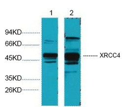
- Western blot analysis of XRCC4 in 1) Hela cells and 2) 293T cells. Samples were probed with a XRCC4 antibody (Product # MA5-24383) at a 1:3000 dilution.

- Submitted by
- Invitrogen Antibodies (provider)
- Main image
- Experimental details
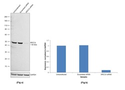
- Knockdown of DNA repair protein XRCC4 was achieved by transfecting U-87 MG with DNA repair protein XRCC4 specific siRNAs (Silencer® select Product # s531571, s531572). Western blot analysis (Fig. a) was performed using Nuclear enriched extracts from the DNA repair protein XRCC4 knockdown cells (lane 3), non-targeting scrambled siRNA transfected cells (lane 2) and untransfected cells (lane 1). The blot was probed with XRCC4 Monoclonal Antibody (5C10) (Product # MA5-24383, 1:2000 ) and Goat anti-Mouse IgG (H+L) Superclonal™ Recombinant Secondary Antibody, HRP (Product # A28177, 1:4000). Densitometric analysis of this western blot is shown in histogram (Fig. b). Decrease in signal upon siRNA mediated knock down confirms that antibody is specific to DNA repair protein XRCC4.

- Submitted by
- Invitrogen Antibodies (provider)
- Main image
- Experimental details
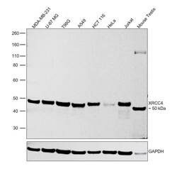
- Western blot was performed using Anti-XRCC4 Monoclonal Antibody (5C10) (Product # MA5-24383) and a 50kDa band corresponding to DNA repair protein XRCC4 was observed. Nuclear enriched extracts (30 µg lysate) of MDA-MB-231 (Lane 1), U-87 MG (Lane 2), T98G (Lane 3), A549 (Lane 4), HCT 116 (Lane 5), HeLa (Lane 6), Jurkat (Lane 7) and tissue extract (30 µg lysate) of Mouse Testis (Lane 8) were electrophoresed using NuPAGE™ 4-12% Bis-Tris Protein Gel (Product # NP0322BOX). Resolved proteins were then transferred onto a Nitrocellulose membrane (Product # IB23001) by iBlot® 2 Dry Blotting System (Product # IB21001). The blot was probed with the primary antibody (1:2000) and detected by chemiluminescence with Goat anti-Mouse IgG (H+L) Superclonal™ Recombinant Secondary Antibody, HRP (Product # A28177, 1:4000) using the iBright FL 1000 (Product # A32752). Chemiluminescent detection was performed using Novex® ECL Chemiluminescent Substrate Reagent Kit (Product # WP20005).

Supportive validation
- Submitted by
- Invitrogen Antibodies (provider)
- Main image
- Experimental details
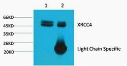
- Immunoprecipitation of XRCC4 in Hela cell lysate using XRCC4 Monoclonal Antibody (5C10) (Product # MA5-24383) at a 1:200 dilution.
