 Explore
 Validate
 Learn
Learn Western blot
 Flow cytometry
Antibody data
- Antibody Data
- Antigen structure
- References [0]
- Comments [0]
- Validations
- Western blot [3]
- Immunocytochemistry [2]
Submit
Validation data
Reference
Comment
Report error
- Product number
- MA5-25112 - Provider product page

- Provider
- Invitrogen Antibodies
- Product name
- HSPB8 Monoclonal Antibody (OTI1E3)
- Antibody type
- Monoclonal
- Antigen
- Recombinant full-length protein
- Reactivity
- Human, Rat
- Host
- Mouse
- Isotype
- IgG
- Antibody clone number
- OTI1E3
- Vial size
- 100 µL
- Concentration
- 0.88 mg/mL
- Storage
- -20° C, Avoid Freeze/Thaw Cycles
No comments: Submit comment
Supportive validation
- Submitted by
- Invitrogen Antibodies (provider)
- Main image

- Experimental details
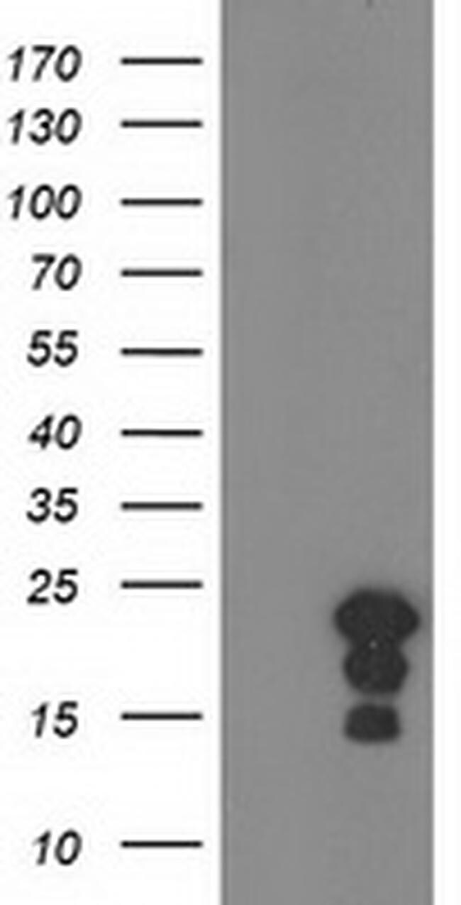
- Western blot analysis of HSPB8 in HEK293T cells in untransfected (Left lane) and transfected (Right lane) samples using 5 µg per lane. The samples were separated by SDS-PAGE and probed with HSPB8 (Product # MA5-25112) monoclonal antibody.
- Submitted by
- Invitrogen Antibodies (provider)
- Main image
- Experimental details
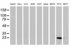
- Western blot analysis of HSPB8 in HepG2, HeLa, SVT2, A549, COS7, Jurkat, MDCK, PC12, MCF7 cells using 35 µg per lane. Samples were probed with HSPB8 (Product # MA5-25112) monoclonal antibody.

- Submitted by
- Invitrogen Antibodies (provider)
- Main image

- Experimental details
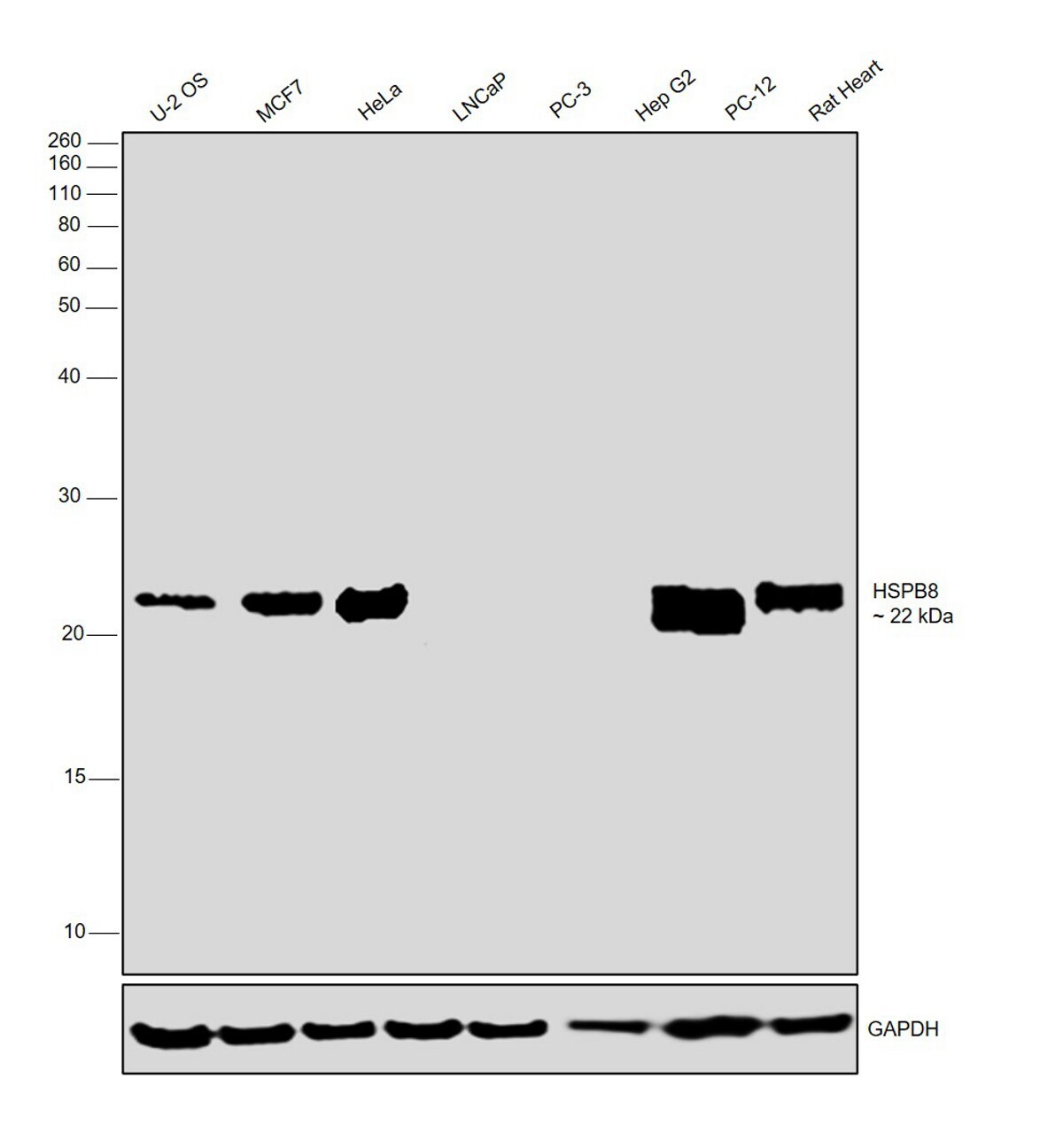
- Western blot was performed using Anti-HSPB8 Monoclonal Antibody (OTI1E3) (Product # MA5-25112) and a 22kDa band corresponding to HSPB8 was observed in cell lines and tissues tested except for LNCaP, PC-3 and Hep G2 which are reported to be low expressing for HSPB8. Whole cell extracts (30 µg lysate) of U-2 OS (Lane 1), MCF7 (Lane 2), HeLa (Lane 3), LNCaP (Lane 4), PC-3 (Lane 5), Hep G2 (Lane 6), PC-12 (Lane 7), Rat Heart (Lane 8) were electrophoresed using NuPAGE™ 4-12% Bis-Tris Protein Gel (Product # NP0321BOX). Resolved proteins were then transferred onto a Nitrocellulose membrane (Product # IB23001) by iBlot® 2 Dry Blotting System (Product # IB21001). The blot was probed with the primary antibody (1:1000 dilution) and detected by chemiluminescence with Goat anti-Mouse IgG (H+L) Superclonal™ Recombinant Secondary Antibody, HRP (Product # A28177, 1:8000 dilution) using the iBright FL 1000 (Product # A32752). Chemiluminescent detection was performed using Novex® ECL Reagent Kit (Product # WP20005).
Supportive validation
- Submitted by
- Invitrogen Antibodies (provider)
- Main image
- Experimental details
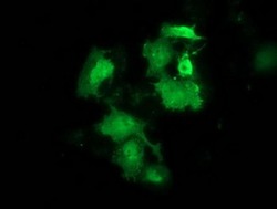
- Immunofluorescent analysis of HSPB8 in COS7 cells. Cells were transfected with a plasmid overexpressing HSPB8 and probed with a HSPB8 monoclonal antibody (Product # MA5-25112).

- Submitted by
- Invitrogen Antibodies (provider)
- Main image
- Experimental details
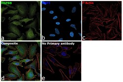
- Immunofluorescence analysis of Heat shock protein beta-8 was performed using 80% confluent log phase HeLa cells. The cells were fixed with 4% paraformaldehyde for 10 minutes, permeabilized with 0.1% Triton™ X-100 for 10 minutes, and blocked with 2% BSA for 10 minutes at room temperature. The cells were labeled with HSPB8 Monoclonal Antibody (OTI1E3) (Product # MA5-25112) at 1:100 dilution in 0.1% BSA, incubated at 4 degree celsius overnight and then labeled with Goat anti-Mouse IgG (H+L) Highly Cross-Adsorbed Secondary Antibody, Alexa Fluor Plus 488 (Product # A32723), (1:2000 dilution), for 45 minutes at room temperature (Panel a: Green). Nuclei (Panel b:Blue) were stained with ProLong™ Diamond Antifade Mountant with DAPI (Product # P36962). F-actin (Panel c: Red) was stained with Rhodamine Phalloidin (Product # R415, 1:300). Panel d represents the merged image showing cytoplasmic and nuclear localization. Panel e represents control cells with no primary antibody to assess background. The images were captured at 60X magnification.
