 Explore
 Validate
 Learn
LearnPA5-17021
antibody from Invitrogen Antibodies
Targeting: DICER1
Dicer, HERNA, K12H4.8-LIKE, KIAA0928, MNG1
 Western blot
 Immunoprecipitation
Antibody data
- Antibody Data
- Antigen structure
- References [0]
- Comments [0]
- Validations
- Western blot [2]
- Immunocytochemistry [1]
Submit
Validation data
Reference
Comment
Report error
- Product number
- PA5-17021 - Provider product page

- Provider
- Invitrogen Antibodies
- Product name
- Dicer Polyclonal Antibody
- Antibody type
- Polyclonal
- Antigen
- Synthetic peptide
- Description
- It is not recommended to aliquot this antibody.
- Reactivity
- Human
- Host
- Rabbit
- Isotype
- IgG
- Vial size
- 100 µL
- Concentration
- 18 µg/mL
- Storage
- -20°C
No comments: Submit comment
Supportive validation
- Submitted by
- Invitrogen Antibodies (provider)
- Main image

- Experimental details
- Knockdown of DICER was achieved by transfecting MCF7 cells with DICER specific siRNAs (Silencer® select Product # s23756). Western blot analysis (Fig. a) was performed using whole cell extracts from DICER knockdown cells (Lane 3), non-specific scrambled siRNA transfected cells (Lane 2) and untransfected cells (Lane 1). The blot was probed with DICER Polyclonal Antibody (Product # PA5-17021, 1:1000 dilution) and Goat anti-Rabbit IgG (H+L) Superclonal™ Recombinant Secondary Antibody, HRP (Product # A27036, 1:4000 dilution). Densitometric analysis of this western blot is shown in histogram (Fig. b). Decrease in signal upon siRNA mediated knock down confirms that antibody is specific to DICER.

- Submitted by
- Invitrogen Antibodies (provider)
- Main image
- Experimental details
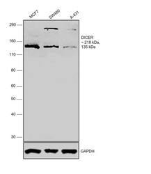
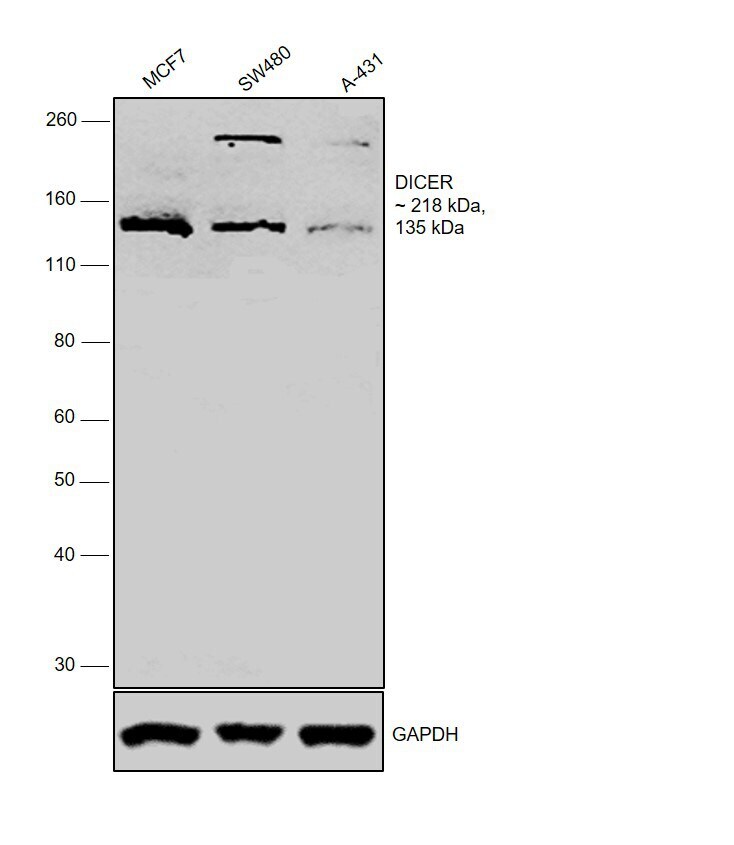
- Western blot was performed using Anti-DICER Polyclonal Antibody (Product # PA5-17021) and ~218 kDa and ~135 kDa bands corresponding to DICER was observed in the cell lines tested. Whole cell extracts (40 µg lysate) of MCF7 (Lane 1), SW480 (Lane 2) and A-431 (Lane 3) were electrophoresed using NuPAGE™ 4-12% Bis-Tris Protein Gel (Product # NP0322BOX). The blot was probed with the primary antibody (1:1000 dilution) and detected by chemiluminescence with Goat anti-Rabbit IgG (H+L), Superclonal™ Recombinant Secondary Antibody, HRP (Product # A27036, 1:4000 dilution) using the iBright FL 1000 (Product # A32752). Chemiluminescent detection was performed using Novex® ECL Chemiluminescent Substrate Reagent Kit (Product # WP20005). Human DICER is a large multidomain protein with two RNase III domains, N-terminal helicase, DUF and PAZ domains. ~135 kDa protein detected could correspond to a C-terminal truncated form of DICER [doi: 10.1016/j.celrep.2013.07.013].
Supportive validation
- Submitted by
- Invitrogen Antibodies (provider)
- Main image

- Experimental details
- Immunofluorescence analysis of DICER was performed using 70% confluent log phase MCF7 cells. The cells were fixed with 4% Paraformaldehyde for 10 minutes, permeabilized with 0.1% Triton™ X-100 for 10 minutes, and blocked with 2% BSA for 10 minutes at room temperature. The cells were labeled with DICER Polyclonal Antibody (Product # PA5-17021) at 1:100 dilution in 0.1% BSA, incubated at 4 degree celsius overnight and then labeled with Goat anti-Rabbit IgG (H+L) Highly Cross-Adsorbed Secondary Antibody, Alexa Fluor Plus 488 (Product # A32731, 1:2000 dilution) for 45 minutes at room temperature (Panel a: Green). Nuclei (Panel b: Blue) were stained with SlowFade® Gold Antifade Mountant with DAPI (Product # S36938). F-actin (Panel c: Red) was stained with Rhodamine Phalloidin (Product # R415, 1:300). Panel d represents the merged image showing cytoplasmic staining of DICER in MCF7. Panel e represents control cells with no primary antibody to assess background. The images were captured at 60X magnification.
