 Explore
 Validate
 Learn
LearnNBP2-29969
antibody from Novus Biologicals
Targeting: RET
CDHF12, CDHR16, HSCR1, MEN2A, MEN2B, MTC1, PTC, RET51
 Western blot
Antibody data
- Antibody Data
- Antigen structure
- References [0]
- Comments [0]
- Validations
- Western blot [2]
- Immunocytochemistry [1]
- Immunohistochemistry [1]
Submit
Validation data
Reference
Comment
Report error
- Product number
- NBP2-29969 - Provider product page

- Provider
- Novus Biologicals
- Product name
- Mouse Monoclonal Ret Antibody
- Antibody type
- Monoclonal
- Description
- Unpurified.
- Reactivity
- Human
- Host
- Mouse
- Isotype
- IgM
- Vial size
- 0.1 ml
- Storage
- Store at 4C short term. Aliquot and store at -20C long term. Avoid freeze-thaw cycles.
No comments: Submit comment
Supportive validation
- Submitted by
- Novus Biologicals (provider)
- Main image

- Experimental details
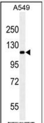
- Western Blot: Ret Antibody (188CT11.2.3) [NBP2-29969] - A549 cell line lysates (35ug/lane).This demonstrates the RET antibody detected the RET protein (arrow).
- Submitted by
- Novus Biologicals (provider)
- Main image
- Experimental details
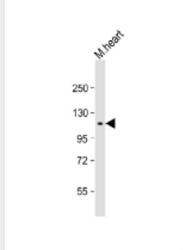
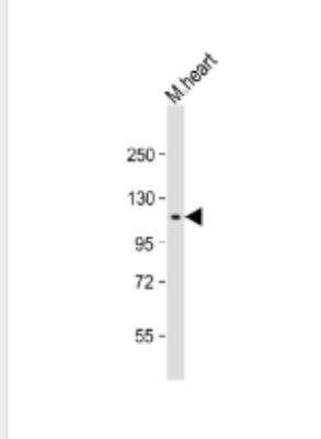
- Western Blot: Ret Antibody (188CT11.2.3) [NBP2-29969] - 1:1000 dilution + mouse heart lysate Secondary Goat Anti-mouse IgM, (H+L),Peroxidase conjugated at 1/10000 dilution. Predicted band size : 124319 Da Blocking/Dilution buffer: 5% NFDM/TBST."
Supportive validation
- Submitted by
- Novus Biologicals (provider)
- Main image

- Experimental details
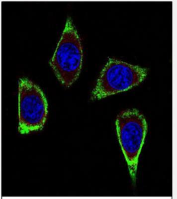
- Immunocytochemistry/Immunofluorescence: Ret Antibody (188CT11.2.3) [NBP2-29969] - RET Antibody (Ascites)(NBP2-29969) with MDA-MB231 cell followed by Alexa Fluor 488-conjugated goat anti-mouse lgG (green).Actin filaments have been labeled with Alexa Fluor? 555 phalloidin (red). DAPI was used to stain the cell nuclear (blue).
Supportive validation
- Submitted by
- Novus Biologicals (provider)
- Main image
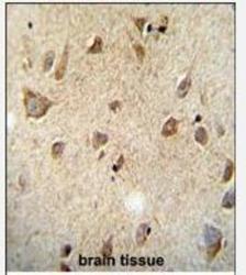
- Experimental details
- Immunohistochemistry-Paraffin: Ret Antibody (188CT11.2.3) [NBP2-29969] - Formalin fixed and paraffin embedded human brain tissue followed by peroxidase conjugation of the secondary antibody and DAB staining. This data demonstrates the use of the RET Monoclonal( Ascites) for immunohistochemistry. Clinical relevance has not been evaluated.
