Explore
 Validate
 Learn
Learn Western blot
Antibody data
- Antibody Data
- Antigen structure
- References [0]
- Comments [0]
- Validations
- Western blot [3]
- Immunocytochemistry [1]
- Immunohistochemistry [2]
Submit
Validation data
Reference
Comment
Report error
- Product number
- MA5-31331 - Provider product page

- Provider
- Invitrogen Antibodies
- Product name
- FABP7 Monoclonal Antibody (CL0236)
- Antibody type
- Monoclonal
- Antigen
- Recombinant full-length protein
- Description
- Immunogen sequence: MVEAFCATWK LTNSQNFDEY MKALGVGFAT RQVGNVTKPT VIISQEGDKV VIRTLSTFKN TEISFQLGEE FDETTADDRN CK
- Reactivity
- Human
- Host
- Mouse
- Isotype
- IgG
- Antibody clone number
- CL0236
- Vial size
- 100 µL
- Concentration
- 1 mg/mL
- Storage
- Store at 4°C short term. For long term storage, store at -20°C, avoiding freeze/thaw cycles.
No comments: Submit comment
Supportive validation
- Submitted by
- Invitrogen Antibodies (provider)
- Main image
- Experimental details
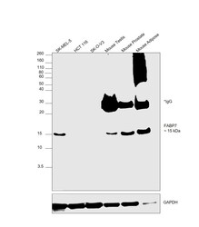
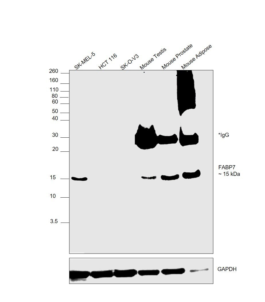
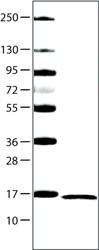
- Western blot was performed using Anti-FABP7 Monoclonal Antibody (CL0236) (Product # MA5-31331) and a 15 kDa band corresponding to Fatty acid-binding protein, brain was observed across cell lines and tissues. Whole cell extracts (40 µg lysate) of SK-MEL-5 (Lane 1), HCT 116 (Lane 2), SK-O-V3 (Lane 3), Mouse Testis (Lane 4), Mouse Prostate (Lane 5), Mouse Adipose (Lane 6) were electrophoresed using NuPAGE™ 12% Bis-Tris Protein Gel (Product # NP0341BOX). Resolved proteins were then transferred onto a nitrocellulose membrane (Product # LC2001) by iBlot® 2 Dry Blotting System (Product # IB21001). The blot was probed with the primary antibody (1:1000 dilution) and detected by chemiluminescence with Goat anti-Mouse IgG (H+L) Superclonal™ Recombinant Secondary Antibody, HRP (Product # A28177,1:20000 dilution) using the iBright FL 1000 (Product # A32752). Chemiluminescent detection was performed using SuperSignal™ West Atto Ultimate Sensitivity Substrate (Product # A38556). HCT116 and SK-O-V3 were low expressing cell models as reported.
- Submitted by
- Invitrogen Antibodies (provider)
- Main image

- Experimental details
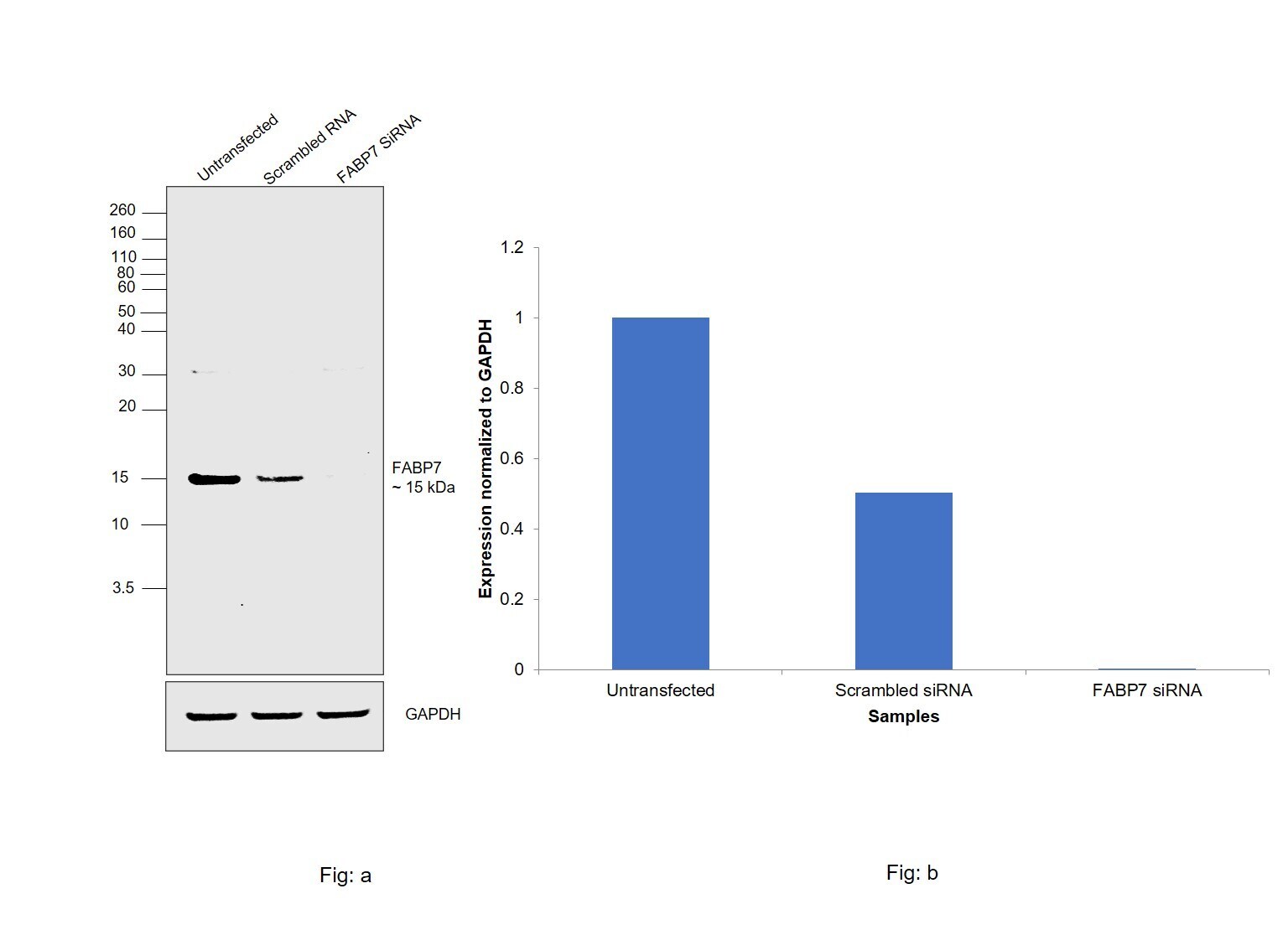
- Knockdown of Fatty acid-binding protein, brain was achieved by transfecting SK-MEL-5 cells with Fatty acid-binding protein, brain specific siRNAs (Silencer® select Product # S223512, S4982). Western blot analysis (Fig. a) was performed using Whole cell extracts from the Fatty acid-binding protein, brain knockdown cells (lane 3), non-targeting scrambled siRNA transfected cells (lane 2) and untransfected cells (lane 1). The blot was probed with FABP7 Monoclonal Antibody (CL0236) (Product # MA5-31331, 1:1000 dilution) and Goat anti-Mouse IgG (H+L) Superclonal™ Recombinant Secondary Antibody, HRP (Product # A28177, 1:20000 dilution). Densitometric analysis of this western blot is shown in histogram (Fig. b). Decrease in signal upon siRNA mediated knock down confirms that antibody is specific to Fatty acid-binding protein, brain.
- Submitted by
- Invitrogen Antibodies (provider)
- Main image
- Experimental details
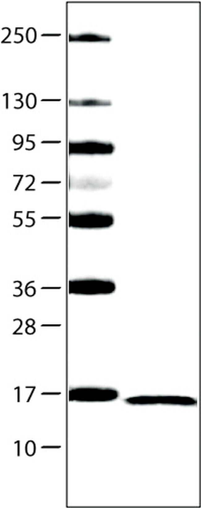
- Western blot analysis of FABP7 by a FABP7 monoclonal antibody (Product # MA5-31331). Lane 1: Marker [kDa] Lane 2: Human cell line U-251 MG.
Supportive validation
- Submitted by
- Invitrogen Antibodies (provider)
- Main image
- Experimental details
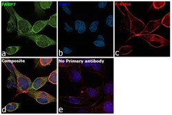
- Immunofluorescence analysis of Fatty acid-binding protein, brain was performed using 70% confluent log phase SK-MEL-5 cells. The cells were fixed with 4% paraformaldehyde for 10 minutes, permeabilized with 0.1% Triton™ X-100 for 15 minutes, and blocked with 2% BSA for 1 hour at room temperature. The cells were labeled with FABP7 Monoclonal Antibody (CL0236) (Product # MA5-31331) at 1:100 dilution in 0.1% BSA, incubated at 4 degree celsius overnight and then labeled with Donkey anti-Mouse IgG (H+L) Highly Cross-Adsorbed Secondary Antibody, Alexa Fluor Plus 488 (Product # A32766), (1:2000 dilution), for 45 minutes at room temperature (Panel a: Green). Nuclei (Panel b:Blue) were stained with ProLong™ Diamond Antifade Mountant with DAPI (Product # P36962). F-actin (Panel c: Red) was stained with Rhodamine Phalloidin (Product # R415, 1:300 dilution). Panel d represents the merged image showing Cytosolic localization. Panel e represents control cells with no primary antibody to assess background. The images were captured at 60X with oil immersion magnification.

Supportive validation
- Submitted by
- Invitrogen Antibodies (provider)
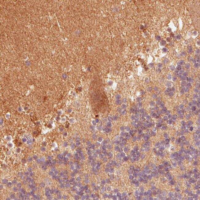
- Main image
- Experimental details
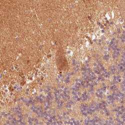
- Immunohistochemical analysis of FABP7 in human cerebellum using a FABP7 monoclonal antibody (Product # MA5-31331). The analysis shows positivity in the molecular cell layer and Purkinje cells.
- Submitted by
- Invitrogen Antibodies (provider)
- Main image
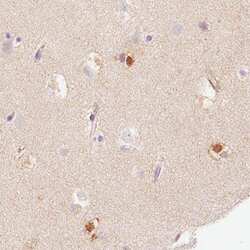
- Experimental details
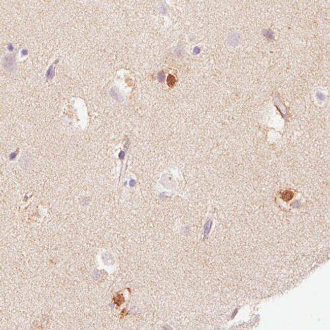
- Immunohistochemical analysis of FABP7 in human cerebral cortex using a FABP7 monoclonal antibody (Product # MA5-31331). The analysis shows immunoreactivity in a subset of glial cells.