Explore
 Validate
 Learn
Learn Western blot
Antibody data
- Antibody Data
- Antigen structure
- References [0]
- Comments [0]
- Validations
- Western blot [5]
- Immunohistochemistry [1]
Submit
Validation data
Reference
Comment
Report error
- Product number
- PA5-21680 - Provider product page

- Provider
- Invitrogen Antibodies
- Product name
- NCKAP1 Polyclonal Antibody
- Antibody type
- Polyclonal
- Antigen
- Recombinant protein fragment
- Description
- Recommended positive controls: NCKAP1-transfected 293T, Mouse brain, rat brain.
- Concentration
- 0.5 mg/mL
No comments: Submit comment
Supportive validation
- Submitted by
- Invitrogen Antibodies (provider)
- Main image

- Experimental details
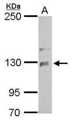
- Western blot analysis of NCKAP1 using 30µg of HepG2 lysate. Samples were loaded onto a 7.5% SDS-PAGE gel and probed with a NCKAP1 polyclonal antibody (Product # PA5-21680) at a dilution of 1:500.
- Submitted by
- Invitrogen Antibodies (provider)
- Main image

- Experimental details
- Western Blot analysis of NCKAP1 was performed by separating 30 µg of non-transfected (–) and transfected (+) 293T whole cell extracts by 5% SDS-PAGE. Proteins were transferred to a membrane and probed with a NCKAP1 Polyclonal Antibody (Product # PA5-21680) at a dilution of 1:5000. The HRP-conjugated anti-rabbit IgG antibody was used to detect the primary antibody.

- Submitted by
- Invitrogen Antibodies (provider)
- Main image

- Experimental details
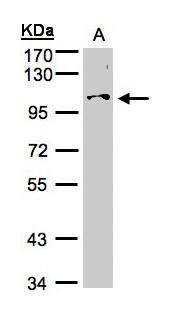
- Western Blot using NCKAP1 Polyclonal Antibody (Product # PA5-21680). Sample (50 µg of whole cell lysate). Lane A: Mouse brain . 5% SDS PAGE. NCKAP1 Polyclonal Antibody (Product # PA5-21680) diluted at 1:1,000.
- Submitted by
- Invitrogen Antibodies (provider)
- Main image
- Experimental details
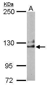
- NCKAP1 Polyclonal Antibody detects NCKAP1 protein by Western blot analysis. A. 50 µg rat brain lysate/extract.5 % SDS-PAGE. NCKAP1 Polyclonal Antibody (Product # PA5-21680) dilution: 1:1,000.

- Submitted by
- Invitrogen Antibodies (provider)
- Main image
- Experimental details
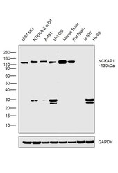
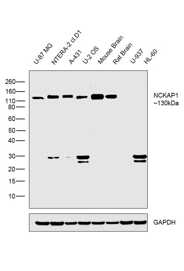
- Western blot was performed using Anti-NCKAP1 Rabbit Polyclonal Antibody (Product # PA5-21680) and a 130kDa band corresponding to NCKAP1 was observed across cell lines and tissues tested except U-937 and HL-60 which are reported negative for NCKP1 expression. An uncharacterized band was also observed at ~30kDa in certain cell lines. Membrane enriched extracts (30 µg lysate) of U-87 MG (Lane 1), NTERA-2 cl.D1 (Lane 2), A-431 (Lane 3), U-2 OS (Lane 4), Mouse Brain (Lane 5), Rat Brain (Lane 6), U-937 (Lane 7) and HL-60 (Lane 8) were electrophoresed using Novex® NuPAGE® 4-12 % Bis-Tris gel (Product # NP0322BOX). Resolved proteins were then transferred onto a nitrocellulose membrane (Product # IB23001) by iBlot® 2 Dry Blotting System (Product # IB21001). The blot was probed with the primary antibody (1:2000 dilution) and detected by chemiluminescence Goat Anti-Rabbit IgG Secondary Antibody, HRP conjugate (Product # A27036, 1:4000 dilution) using the iBright FL 1000 (Product # A32752). Chemiluminescent detection was performed using Novex® ECL Chemiluminescent Substrate Reagent Kit (Product # WP20005).
Supportive validation
- Submitted by
- Invitrogen Antibodies (provider)
- Main image
- Experimental details
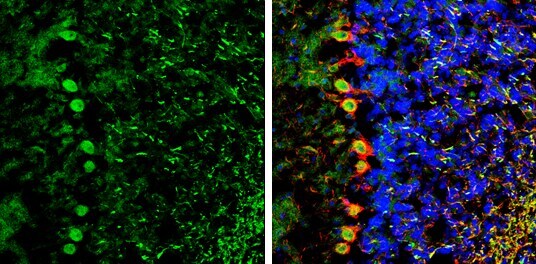
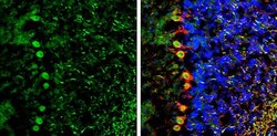
- Immunohistochemistry (Frozen) analysis of NCKAP1 was performed in frozen-sectioned mouse cerebellum tissue using NCKAP1 Polyclonal Antibody (Product # PA5-21680) at a dilution of 1:250 (Green). Red: NF-H, stained by NF-H antibody diluted at 1:500. Blue: Fluoroshield with DAPI.