 Explore
 Validate
 Learn
LearnNBP1-88606
antibody from Novus Biologicals
Targeting: TGFBI
BIGH3, CDB1, CDGG1, CSD1, CSD2, CSD3, LCD1
 Western blot
Antibody data
- Antibody Data
- Antigen structure
- References [4]
- Comments [0]
- Validations
- Western blot [2]
- Immunohistochemistry [14]
Submit
Validation data
Reference
Comment
Report error
- Product number
- NBP1-88606 - Provider product page

- Provider
- Novus Biologicals
- Proper citation
- Novus Cat#NBP1-88606, RRID:AB_11027801
- Product name
- Rabbit Polyclonal Beta Ig-h3/TGFBI Antibody
- Antibody type
- Polyclonal
- Description
- Immunogen affinity purified. Specificity of human Beta Ig-h3/TGFBI antibody verified on a Protein Array containing target protein plus 383 other non-specific proteins.
- Reactivity
- Human
- Host
- Rabbit
- Isotype
- IgG
- Vial size
- 0.1 ml
- Storage
- Store at 4C short term. Aliquot and store at -20C long term. Avoid freeze-thaw cycles.
Submitted references TGFBI protein high expression predicts poor prognosis in colorectal cancer patients.
Association of TCF4 and CLU polymorphisms with Fuchs' endothelial dystrophy and implication of CLU and TGFBI proteins in the disease process.
SILAC-based quantitative proteomic approach to identify potential biomarkers from the esophageal squamous cell carcinoma secretome.
SILAC-based quantitative proteomic approach to identify potential biomarkers from the esophageal squamous cell carcinoma secretome.
Zhu J, Chen X, Liao Z, He C, Hu X
International journal of clinical and experimental pathology 2015;8(1):702-10
International journal of clinical and experimental pathology 2015;8(1):702-10
Association of TCF4 and CLU polymorphisms with Fuchs' endothelial dystrophy and implication of CLU and TGFBI proteins in the disease process.
Kuot A, Hewitt AW, Griggs K, Klebe S, Mills R, Jhanji V, Craig JE, Sharma S, Burdon KP
European journal of human genetics : EJHG 2012 Jun;20(6):632-8
European journal of human genetics : EJHG 2012 Jun;20(6):632-8
SILAC-based quantitative proteomic approach to identify potential biomarkers from the esophageal squamous cell carcinoma secretome.
Kashyap MK, Harsha HC, Renuse S, Pawar H, Sahasrabuddhe NA, Kim MS, Marimuthu A, Keerthikumar S, Muthusamy B, Kandasamy K, Subbannayya Y, Prasad TS, Mahmood R, Chaerkady R, Meltzer SJ, Kumar RV, Rustgi AK, Pandey A
Cancer biology & therapy 2010 Oct 15;10(8):796-810
Cancer biology & therapy 2010 Oct 15;10(8):796-810
No comments: Submit comment
Supportive validation
- Submitted by
- Novus Biologicals (provider)
- Main image

- Experimental details
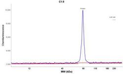
- Simple Western: Beta Ig-h3/TGFBI Antibody [NBP1-88606] - Simple Western lane view shows a specific band for Beta Ig-h3/TGFBI in 0.2 mg/ml of U-251 MG lysate(s). This experiment was performed under reducing conditions using the 12-230 kDa separation systems.

- Submitted by
- Novus Biologicals (provider)
- Main image
- Experimental details
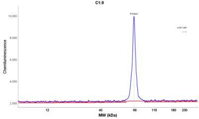
- Simple Western: Beta Ig-h3/TGFBI Antibody [NBP1-88606] - Electropherogram image of the corresponding Simple Western lane view. Beta Ig-h3/TGFBI antibody was used at 1:25 dilution on U-251 MG lysate(s) respectively.
Supportive validation
- Submitted by
- Novus Biologicals (provider)
- Main image
- Experimental details
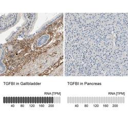
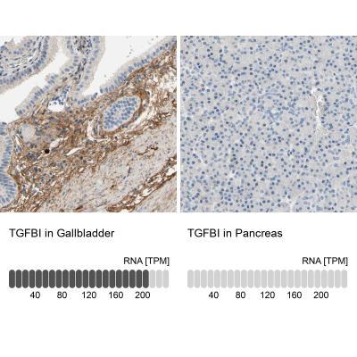
- Immunohistochemistry-Paraffin: Beta Ig-h3/TGFBI Antibody [NBP1-88606] - Immunohistochemistry analysis in human gallbladder and pancreas tissues. Corresponding RNA-seq data are presented for the same tissues.
- Submitted by
- Novus Biologicals (provider)
- Main image

- Experimental details
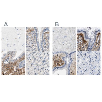
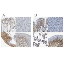
- Immunohistochemistry-Paraffin: Beta Ig-h3/TGFBI Antibody [NBP1-88606] - Staining of human cerebral cortex, colon, gallbladder and stomach using Anti-TGFBI antibody NBP1-88606 (A) shows similar protein distribution across tissues to independent antibody NBP2-48534 (B).
- Submitted by
- Novus Biologicals (provider)
- Main image
- Experimental details
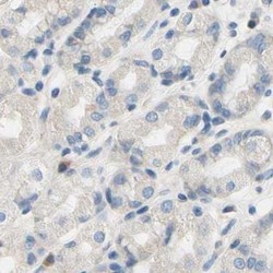
- Immunohistochemistry-Paraffin: Beta Ig-h3/TGFBI Antibody [NBP1-88606] - Staining of human stomach.
- Submitted by
- Novus Biologicals (provider)
- Main image
- Experimental details
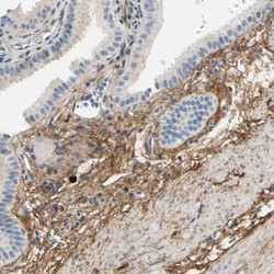
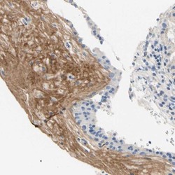
- Immunohistochemistry-Paraffin: Beta Ig-h3/TGFBI Antibody [NBP1-88606] - Staining of human gallbladder shows moderate to strong cytoplasmic positivity in stromal cells.

- Submitted by
- Novus Biologicals (provider)
- Main image
- Experimental details
- Immunohistochemistry-Paraffin: Beta Ig-h3/TGFBI Antibody [NBP1-88606] - Staining of human placenta shows moderate to strong membranous positivity in stromal cells.

- Submitted by
- Novus Biologicals (provider)
- Main image

- Experimental details
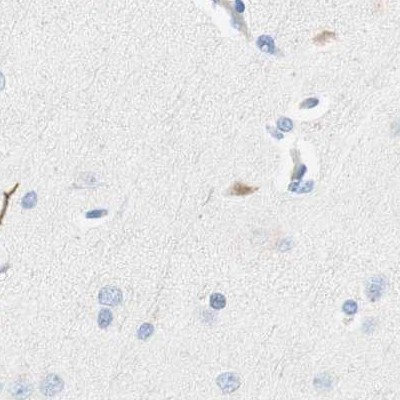
- Immunohistochemistry-Paraffin: Beta Ig-h3/TGFBI Antibody [NBP1-88606] - Staining of human cerebral cortex.
- Submitted by
- Novus Biologicals (provider)
- Main image

- Experimental details
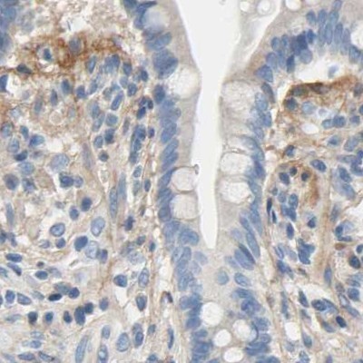
- Immunohistochemistry-Paraffin: Beta Ig-h3/TGFBI Antibody [NBP1-88606] - Staining of human colon.
- Submitted by
- Novus Biologicals (provider)
- Main image

- Experimental details
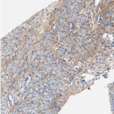
- Immunohistochemistry-Paraffin: Beta Ig-h3/TGFBI Antibody [NBP1-88606] - Staining of human endometrium shows moderate to strong membranous positivity in stromal cells.
- Submitted by
- Novus Biologicals (provider)
- Main image

- Experimental details
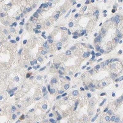
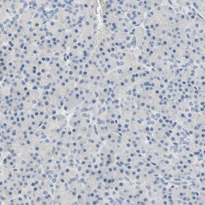
- Immunohistochemistry-Paraffin: Beta Ig-h3/TGFBI Antibody [NBP1-88606] - Staining of human pancreas shows no positivity in exocrine glandular cells as expected.

- Submitted by
- Novus Biologicals (provider)
- Main image
- Experimental details
- Immunohistochemistry-Paraffin: Beta Ig-h3/TGFBI Antibody [NBP1-88606] - Staining of human gastrointestinal, pancreas, placenta and skin using Anti-Beta Ig-h3/TGFBI antibody NBP1-88606 (A) shows similar protein distribution across tissues to independent antibody NBP2-48534 (B).

- Submitted by
- Novus Biologicals (provider)
- Main image
- Experimental details
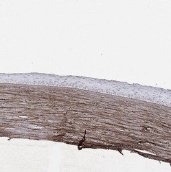
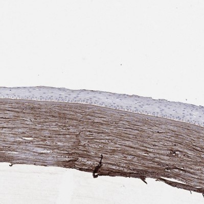
- Immunohistochemistry-Paraffin: Beta Ig-h3/TGFBI Antibody [NBP1-88606] - Staining of human eye shows moderate extracellular positivity in cornea.
- Submitted by
- Novus Biologicals (provider)
- Main image
- Experimental details
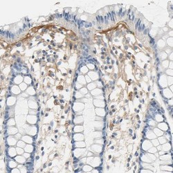
- Immunohistochemistry-Paraffin: Beta Ig-h3/TGFBI Antibody [NBP1-88606] - Staining of human gastrointestinal shows moderate positivity in extracellular matrix of glandular cells.

- Submitted by
- Novus Biologicals (provider)
- Main image

- Experimental details
- Immunohistochemistry-Paraffin: Beta Ig-h3/TGFBI Antibody [NBP1-88606] - Staining of human pancreas shows no positivity in exocrine glandular cells as expected.
- Submitted by
- Novus Biologicals (provider)
- Main image

- Experimental details
- Immunohistochemistry-Paraffin: Beta Ig-h3/TGFBI Antibody [NBP1-88606] - Staining of human skin shows moderate positivity in extracellular matrix of dermis.
