 Explore
 Validate
 Learn
Learn17-1529-41
antibody from Invitrogen Antibodies
Targeting: CTLA4
CD, CD152, CELIAC3, CTLA-4, GSE, IDDM12
 Flow cytometry
Antibody data
- Antibody Data
- Antigen structure
- References [6]
- Comments [0]
- Validations
- Flow cytometry [1]
- Other assay [4]
Submit
Validation data
Reference
Comment
Report error
- Product number
- 17-1529-41 - Provider product page

- Provider
- Invitrogen Antibodies
- Product name
- CD152 (CTLA-4) Monoclonal Antibody (14D3), APC, eBioscience™
- Antibody type
- Monoclonal
- Antigen
- Other
- Description
- Description: The 14D3 monoclonal antibody reacts with human CD152, also known as cytotoxic T lymphocyte antigen-4 (CTLA-4). CTLA-4, a protein with structural similarities to CD28, is expressed on activated T cells (activated B cells may also express CTLA-4) and binds the B7 family members, CD80 (B7-1) and CD86 (B7-2), with higher affinity than CD28 does. CTLA-4 and CD28 appear to deliver opposing signals to T cells: while CD28 is a potent costimulator, CTLA-4 restricts the progression of T cells to an activated state by inhibiting IL-2 secretion and cellular proliferation. The cytoplasmic portion of CTLA-4 contains ER retention motifs, resulting in intracellular localization of a large proportion of newly synthesized CTLA-4 in response to TCR signaling.
- Antibody clone number
- 14D3
- Concentration
- 5 µL/Test
Submitted references Cancer stem-like cells evade CD8(+)CD103(+) tumor-resident memory T (T(RM)) lymphocytes by initiating an epithelial-to-mesenchymal transition program in a human lung tumor model.
Engineering advanced logic and distributed computing in human CAR immune cells.
Methylome-based cell-of-origin modeling (Methyl-COOM) identifies aberrant expression of immune regulatory molecules in CLL.
CD19-targeted CAR regulatory T cells suppress B cell pathology without GvHD.
Accumulation of TNFR2-expressing regulatory T cells in malignant pleural effusion of lung cancer patients is associated with poor prognosis.
A cellular platform for the evaluation of immune checkpoint molecules.
Corgnac S, Damei I, Gros G, Caidi A, Terry S, Chouaib S, Deloger M, Mami-Chouaib F
Journal for immunotherapy of cancer 2022 Apr;10(4)
Journal for immunotherapy of cancer 2022 Apr;10(4)
Engineering advanced logic and distributed computing in human CAR immune cells.
Cho JH, Okuma A, Sofjan K, Lee S, Collins JJ, Wong WW
Nature communications 2021 Feb 4;12(1):792
Nature communications 2021 Feb 4;12(1):792
Methylome-based cell-of-origin modeling (Methyl-COOM) identifies aberrant expression of immune regulatory molecules in CLL.
Wierzbinska JA, Toth R, Ishaque N, Rippe K, Mallm JP, Klett LC, Mertens D, Zenz T, Hielscher T, Seifert M, Küppers R, Assenov Y, Lutsik P, Stilgenbauer S, Roessner PM, Seiffert M, Byrd J, Oakes CC, Plass C, Lipka DB
Genome medicine 2020 Mar 18;12(1):29
Genome medicine 2020 Mar 18;12(1):29
CD19-targeted CAR regulatory T cells suppress B cell pathology without GvHD.
Imura Y, Ando M, Kondo T, Ito M, Yoshimura A
JCI insight 2020 Jul 23;5(14)
JCI insight 2020 Jul 23;5(14)
Accumulation of TNFR2-expressing regulatory T cells in malignant pleural effusion of lung cancer patients is associated with poor prognosis.
Ye LL, Peng WB, Niu YR, Xiang X, Wei XS, Wang ZH, Wang X, Zhang SY, Chen X, Zhou Q
Annals of translational medicine 2020 Dec;8(24):1647
Annals of translational medicine 2020 Dec;8(24):1647
A cellular platform for the evaluation of immune checkpoint molecules.
Jutz S, Hennig A, Paster W, Asrak Ö, Dijanovic D, Kellner F, Pickl WF, Huppa JB, Leitner J, Steinberger P
Oncotarget 2017 Sep 12;8(39):64892-64906
Oncotarget 2017 Sep 12;8(39):64892-64906
No comments: Submit comment
Supportive validation
- Submitted by
- Invitrogen Antibodies (provider)
- Main image

- Experimental details
- Unstimulated (blue histogram) or 3-day PHA (Product # 00-4977-03)-stimulated normal human peripheral blood cells (purple histogram) were intracellularly stained with Anti-Human CD152 (CTLA-4) APC using the Intracellular Fixation and Permeabilization Buffer Set (Product # 88-8824-00) and protocol. Viable cells in the lymphocyte gate, as determined by Fixable Viability Dye eFluor® 450 (Product # 65-0863-14), were used for analysis.

Supportive validation
- Submitted by
- Invitrogen Antibodies (provider)
- Main image
- Experimental details
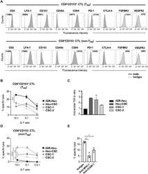
- Resident memory T (T RM ) and non-T RM phenotypic profiles and susceptibility of cancer stem cell (CSC) to cytotoxic T lymphocyte (CTL)-mediated killing. (A) Flow cytometry analyses of CD8, LFA-1, CD103, CD49a, CD69, PD-1, CTLA-4, TGFBR2 and VEGFR2 on CD8 + CD103 + T RM (Heu171) and CD8 + CD103 - non-T RM (H32-22) clones. Mean immunofluorescence intensity (MFI) are in parentheses. (B) Cytotoxic activity of the T RM clone (Heu171) towards autologous IGR-Heu, Heu-CSC, CSC-1 and CSC-2 target cells. Percent of specific lysis are shown at indicated effector to target (E:T) ratios. (C) Quantification of transforming growth factor (TGF)-beta in conditioned media from IGR-Heu, Heu-CSC, CSC-1 and CSC-2 by multi-analyte flow assay. Ratios of active/total TGF-beta normalized to IGR-Heu are included. Results are presented as mean+-SEM of duplicates. (D) Cytotoxicity of the non-T RM clone (H32-22) towards autologous IGR-Heu, Heu-CSC, CSC-1 and CSC-2 target cells. (E) Inhibition of T RM -cell-mediated killing. CSC-1 cells are preincubated in the presence of isotype control, anti-major histocompatibility complex class I (MHC-I) or anti-intercellular adhesion molecule 1 (ICAM-1) neutralizing monoclonal antibody (mAb) and then CTL were added at 5:1 E:T ratio. Symbols represent replicates and horizontal bars represent means+-SEM (n=3). P value was determined by two-way analysis of variance test. *p
- Submitted by
- Invitrogen Antibodies (provider)
- Main image

- Experimental details
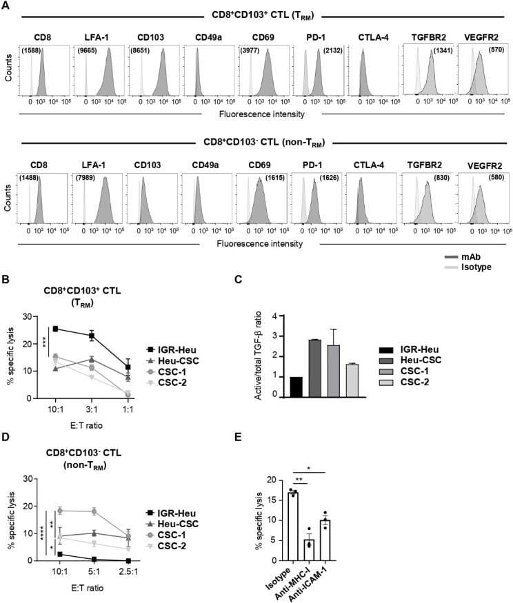
- Effects of TNF on the suppressive function of TNFR2 + Tregs. Purified CD4 + T cells were cultured in medium alone, TNF (50 ng/mL), TNF combined with anti-TNFR2 mAbs (10 ug/mL), or isotype IgG, as indicated, for 72 hours. The representative FACS analysis of (A) CTLA-4 + cells and (B) PD-L1 + cells in TNFR2 + Tregs as indicated, gated on live CD4 + Foxp3 + cells. Summary of the proportions of (C) CTLA-4 + cells (n=3) and (D) PD-L1 + cells (n=3) in Tregs within each condition. Data are expressed as means +- SEM. *, P
- Submitted by
- Invitrogen Antibodies (provider)
- Main image
- Experimental details
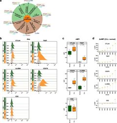
- Fig. 6 Flow cytometry analysis of T cell-/lymphocyte-specific markers on normal and malignant B cells from CLL patients. a Summary scheme representing functional implications of CLL-specific candidate genes selected for flow cytometric analysis. b Flow cytometric analysis of expression of CTLA-4, TIGIT, CD276, LILRB4, and CD2 on peripheral blood B cells of CLL patients. The expression was determined for non-malignant B cells (""Normal""; CD19 + CD5 - B cells, represented in green) and neoplastic B cells (""CLL"", CD19 + CD5 + B cells, represented in orange) detected in the same samples. ""Co,"" no antibody staining control; ""Ab,"" staining with the antibody of interest as indicated. c Normalized median fluorescence intensities (target MFI - MFI of negative control [Co]; nMFI). d Delta normalized median fluorescence intensities between CLL cells and normal B cells (DeltanMFI (CLL-normal)) for each patient tested

- Submitted by
- Invitrogen Antibodies (provider)
- Main image

- Experimental details
- Fig. 3 The intracellular AND logic with different signaling domains. a Diagram of intracellular AND logic. b Primary human CD8+ T cells were transduced with FOS zipCAR-containing CD3zeta domain and RR zipCAR-containing CD28 domain. Cytotoxicity against Her2- and Axl-expressing Nalm6 was measured 24 h after adding alpha-Her2-SYN9 and/or alpha-Axl-EE zipFvs. The heatmap indicates cytotoxicity at varying zipFv concentrations ( n = 3, data are represented as mean). c Cytotoxicity of CD8+ T cells transduced with FOS zipCAR-containing CD3zeta domain and RR zipCAR-containing 4-1BB domain. The heatmap indicates cytotoxicity at varying zipFv concentrations ( n = 3, data are represented as mean). d (Left) Isolated Treg cells were transduced with two zipCAR constructs: SYN6-CD3zeta-P2A-FOXP3 and SYN1-CD28-P2A-puro. After puromycin selection (2 mug/mL), Treg cells were co-cultured with Her2- and Axl-expressing Nalm6 target cells (Right) The heatmap shows surface CTLA-4 expression detected after 48 h by flow cytometry at varying zipFv concentrations (alpha-Axl-SYN5 and alpha-Her2-SYN2) ( n = 3, data are represented as mean).
