Explore
 Validate
 Learn
Learn Western blot
Antibody data
- Antibody Data
- Antigen structure
- References [0]
- Comments [0]
- Validations
- Western blot [1]
- Flow cytometry [1]
- Blocking/Neutralizing [1]
Submit
Validation data
Reference
Comment
Report error
- Product number
- MAB10861 - Provider product page

- Provider
- R&D Systems
- Product name
- Human PD-1 Antibody
- Antibody type
- Monoclonal
- Description
- Protein A or G purified from cell culture supernatant. Detects human PD-1 in direct ELISAs.
- Reactivity
- Human
- Host
- Mouse
- Conjugate
- Unconjugated
- Antigen sequence
Q15116- Isotype
- IgG
- Antibody clone number
- 913429
- Vial size
- 100 ug
- Storage
- Use a manual defrost freezer and avoid repeated freeze-thaw cycles. 12 months from date of receipt, -20 to -70 °C as supplied. 1 month, 2 to 8 °C under sterile conditions after reconstitution. 6 months, -20 to -70 °C under sterile conditions after reconstitution.
No comments: Submit comment
Supportive validation
- Submitted by
- R&D Systems (provider)
- Main image
- Experimental details
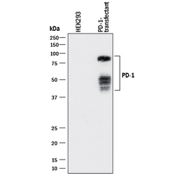
- Detection of Human PD-1 by Western Blot. Western blot shows lysates of HEK293 human embryonic kidney cell line either mock transfected or transfected with human PD-1. PVDF membrane was probed with 1 µg/mL of Mouse Anti-Human PD-1 Monoclonal Antibody (Catalog # MAB10861) followed by HRP-conjugated Anti-Mouse IgG Secondary Antibody (Catalog # HAF018). Specific bands were detected for PD-1 at approximately 40-80 kDa (as indicated). This experiment was conducted under reducing conditions and using Immunoblot Buffer Group 1.

Supportive validation
- Submitted by
- R&D Systems (provider)
- Main image
- Experimental details
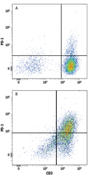
- Detection of PD-1 in Human PBMCs by Flow Cytometry. Human peripheral blood mononuclear cells (PBMCs) either (A) untreated or (B) treated with 5 ng/mL PHA for 2 days were stained with Mouse Anti-Human PD-1 Monoclonal Antibody (Catalog # MAB10861) followed by Allophycocyanin-conjugated Anti-Mouse IgG Secondary Antibody (Catalog # F0101B) and Mouse Anti-Human CD3 epsilon PE-conjugated Monoclonal Antibody (Catalog # FAB100P). Quadrant markers were set based on control antibody staining (Catalog # MAB002).

Supportive validation
- Submitted by
- R&D Systems (provider)
- Main image

- Experimental details
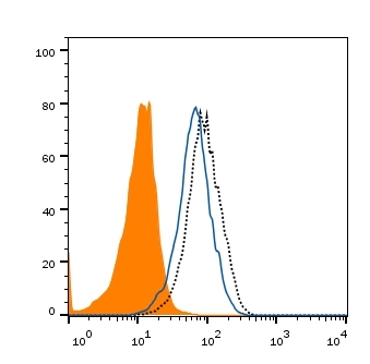
- PD-1 Binding to B7-H1-transfected HEK293 Human Cell Line is Blocked by Human PD-1 Antibody. In a functional flow cytometry test, biotinylated recombinant human B7-H1 (10 ng/mL, Catalog # 9049-B7) binds to HEK293 human embryonic kidney cell line transfected with human PD-1 (black dotted line). Binding is completely blocked (orange histogram) by 2.5 μg/mL of Mouse Anti-Human PD-1 Monoclonal Antibody (Catalog # MAB10861). Mouse IgG2B Isotype Control (Catalog # MAB004) at 2.5 μg/mL was used as a control (blue line). Cells were stained with Streptavidin-APC (Catalog # F0050).