Explore
 Validate
 Learn
Learn Western blot
 Immunocytochemistry
Antibody data
- Antibody Data
- Antigen structure
- References [0]
- Comments [0]
- Validations
- Western blot [1]
- Immunohistochemistry [1]
- Flow cytometry [1]
Submit
Validation data
Reference
Comment
Report error
- Product number
- NBP2-76329 - Provider product page

- Provider
- Novus Biologicals
- Product name
- Rabbit Monoclonal ATF6 Antibody
- Antibody type
- Monoclonal
- Description
- Protein A or G purified.
- Reactivity
- Human, Mouse, Rat
- Host
- Rabbit
- Isotype
- IgG
- Vial size
- 0.1 mg
- Concentration
- 1.0 mg/ml
- Storage
- Store at 4C for up to 3 months. For longer storage, aliquot and store at -20C.
No comments: Submit comment
Supportive validation
- Submitted by
- Novus Biologicals (provider)
- Main image
- Experimental details
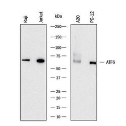
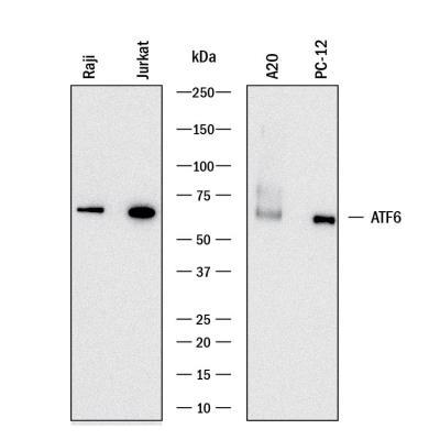
- Western Blot: ATF6 Antibody [NBP2-76329] - Western blot shows lysates of Raji human Burkitt's lymphoma cell line, Jurkat human acute T cell leukemia cell line, A20 mouse B cell lymphoma cell line, and PC-12 rat adrenal pheochromocytoma cell line. PVDF membrane was probed with 0.05 ug/mL of Rabbit Anti-Human/Mouse/Rat ATF6 Monoclonal Antibody (Catalog # NBP2-76329) followed by HRP-conjugated Anti-Rabbit IgG Secondary Antibody (Catalog # HAF008). A specific band was detected for ATF6 at approximately 70 kDa (as indicated). This experiment was conducted under reducing conditions.
Supportive validation
- Submitted by
- Novus Biologicals (provider)
- Main image

- Experimental details
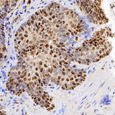
- Immunohistochemistry: ATF6 Antibody (2358C) [NBP2-76329] - ATF6 was detected in immersion fixed paraffin-embedded sections of human prostate using Rabbit Anti-Human/Mouse/Rat ATF6 Monoclonal Antibody (Catalog # NBP2-76329) at 0.3 ug/mL for 1 hour at room temperature followed by incubation with the Anti-Rabbit IgG VisUCyte(TM) HRP Polymer Antibody (Catalog # VC003). Before incubation with the primary antibody, tissue was subjected to heat-induced epitope retrieval using Antigen Retrieval Reagent-Basic (Catalog # CTS013). Tissue was stained using DAB (brown) and counterstained with hematoxylin (blue). Specific staining was localized to cytoplasm and nuclei.
Supportive validation
- Submitted by
- Novus Biologicals (provider)
- Main image

- Experimental details
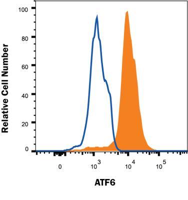
- Flow Cytometry: ATF6 Antibody [NBP2-76329] - MCF-7 human breast cancer cell line was stained with Rabbit Anti-Human/Mouse/Rat ATF6 Monoclonal Antibody (Catalog # NBP2-76329, filled histogram) or Rabbit IgG control antibody (Catalog # MAB1050, open histogram) followed by APC-conjugated Anti-Rabbit IgG Secondary Antibody (Catalog # F0111). To facilitate intracellular staining, cells were fixed and permeabilized with FlowX FoxP3 Fixation & Permeabilization Buffer Kit (Catalog # FC012).