Explore
 Validate
 Learn
Learn Western blot
Antibody data
- Antibody Data
- Antigen structure
- References [0]
- Comments [0]
- Validations
- Western blot [1]
- Immunocytochemistry [2]
Submit
Validation data
Reference
Comment
Report error
- Product number
- 711777 - Provider product page

- Provider
- Invitrogen Antibodies
- Product name
- HK1 Recombinant Polyclonal Antibody
- Antibody type
- Polyclonal
- Antigen
- Synthetic peptide
- Description
- This antibody is predicted to react with Monkey, Horse, Dog, Bovine.
- Concentration
- 0.5 mg/mL
No comments: Submit comment
Supportive validation
- Submitted by
- Invitrogen Antibodies (provider)
- Main image
- Experimental details
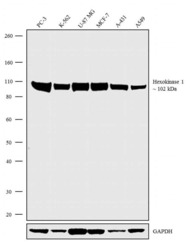
- Western blot analysis was performed on Whole cell extracts (60 µg lysate) of PC-3 (Lane 1), K-562 (Lane 2), U-87 MG (Lane 3), MCF-7 (Lane 4), A-431 (Lane 5) and A549 (Lane 6). The blots were probed with Anti-Hexokinase 1 Recombinant Rabbit Polyclonal Antibody (Product # 711777, 2.5 µg/mL) and detected by chemiluminescence using Goat anti-Rabbit IgG (H+L) Superclonal™ Secondary Antibody, HRP conjugate (Product # A27036, 0.25 µg/mL, 1:4000 dilution). A 102 kDa band corresponding to Hexokinase 1 was observed across the cell lines tested. Known quantity of protein samples were electrophoresed using Novex®NuPAGE®4-12% Bis-Tris gel (Product # NP0322BOX), XCell SureLock™ Electrophoresis System (Product # EI0002) and Novex® Sharp Pre-Stained Protein Standard (Product # LC5800). Resolved proteins were then transferred onto a nitrocellulose membrane with iBlot® Dry Blotting System (Product # IB21001). The membrane was probed with the relevant primary and secondary Antibody following blocking with 5% skimmed milk. Chemiluminescent detection was performed using Pierce™ ECL Western blotting Substrate (Product # 32106).

Supportive validation
- Submitted by
- Invitrogen Antibodies (provider)
- Main image
- Experimental details
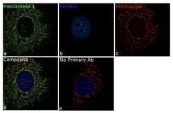
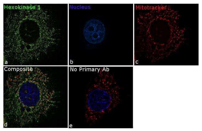
- For immunofluorescence analysis, MCF7 cells were fixed and permeabilized for detection of endogenous Hexokinase-1 using Anti-Hexokinase-1 Recombinant Rabbit Polyclonal Antibody (Product # 711777, 5 µg/mL) and labeled with Goat anti-Rabbit IgG (H+L) Superclonal™ Secondary Antibody, Alexa Fluor® 488 conjugate (Product # A27034, 1:2000). Panel a) shows representative cells that were stained for detection and localization of Hexokinase-1 protein (green), Panel b) is stained for nuclei (blue) using SlowFade® Gold Antifade Mountant with DAPI (Product # S36938). Panel c) represents mitochondrial staining using MitoTracker® Red CMXRos (Product # M7512). Panel d) is a composite image of Panels a, b and c clearly demonstrating co-localization of Hexokinase-1 with mitotracker which specifically binds to the mitochondria. Panel e) represents control cells without primary antibody to assess background. The images were captured at 60X magnification.
- Submitted by
- Invitrogen Antibodies (provider)
- Main image
- Experimental details
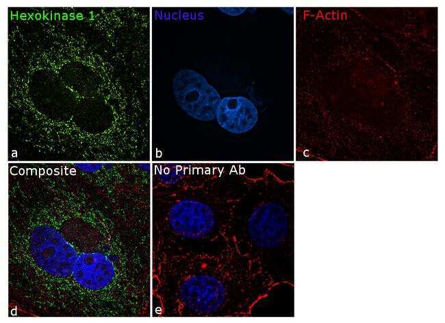
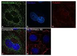
- For immunofluorescence analysis, MCF7 cells were fixed and permeabilized for detection of endogenous Hexokinase-1 using Anti- Hexokinase-1 Recombinant Rabbit Polyclonal Antibody (Product # 711777, 5 µg/mL) and labeled with Goat anti-Rabbit IgG (H+L) Superclonal™ Secondary Antibody, Alexa Fluor® 488 conjugate (Product # A27034, 1:2000). Panel a) shows representative cells that were stained for detection and localization of Hexokinase-1 protein (green), Panel b) is stained for nuclei (blue) using SlowFade® Gold Antifade Mountant with DAPI (Product # S36938). Panel c) represents cytoskeletal F-actin staining using Rhodamine Phalloidin (Product # R415, 1:300). Panel d) is a composite image of Panels a, b and c clearly demonstrating mitochondrial localization of Hexokinase-1. Panel e) represents control cells with no primary antibody to assess background. The images were captured at 60X magnification.