Explore
 Validate
 Learn
Learn Western blot
Antibody data
- Antibody Data
- Antigen structure
- References [0]
- Comments [0]
- Validations
- Western blot [2]
Submit
Validation data
Reference
Comment
Report error
- Product number
- 702266 - Provider product page

- Provider
- Invitrogen Antibodies
- Product name
- Phospho-RSK1 (Ser380) Recombinant Rabbit Monoclonal Antibody (13H23L1)
- Antibody type
- Monoclonal
- Antigen
- Synthetic peptide
- Description
- This antibody is predicted to react with Monkey, Bovine, Mouse, Rat
- Antibody clone number
- 13H23L1
- Concentration
- 0.5 mg/mL
No comments: Submit comment
Supportive validation
- Submitted by
- Invitrogen Antibodies (provider)
- Main image

- Experimental details
- Western blot analysis was performed on Whole cell extracts (30 µg lysate) of HeLa, HeLa (Serum starved for overnight and treated with 25 ng/mL of TNFa for 20 min). The blots were probed with Anti-Phospho-RSK1 (Ser380) Recombinant Rabbit Monoclonal Antibody (Product # 702266, 1-2 µg/mL) (Lane 1, 2). To confirm the specificity of Phospho-RSK1 (Ser380), competition was performed with the phospopeptide (10 µg/mL) (Lane 3, 4) and non phospopeptide (10 µg/mL) (Lane 5, 6). A 90 kDa band corresponding to RSK1 (pS380) was observed in HeLa (with increase in expression upon treatment) when probed with the antibody alone or with non-phospho peptide competition. The blots were detected by chemiluminescence using Goat anti-Rabbit IgG (H+L) Superclonal Secondary Antibody, HRP conjugate (Product # A27036, 0.4 µg/mL, 1:2500 dilution). Known quantity of protein samples were electrophoresed using Novex® NuPAGE® 4-12% Bis-Tris gel (Product # NP0321BOX), XCell SureLock Electrophoresis System (Product # EI0002) and Novex® Sharp Pre-Stained Protein Standard (Product # LC5800). Resolved proteins were then transferred onto a nitrocellulose membrane with iBlot® Dry Blotting System (Product # IB21001). The membrane was probed with the relevant primary and secondary Antibody following blocking with 5% skimmed milk. Chemiluminescent detection was performed using Pierce™ ECL Western blotting Substrate (Product # 32106).

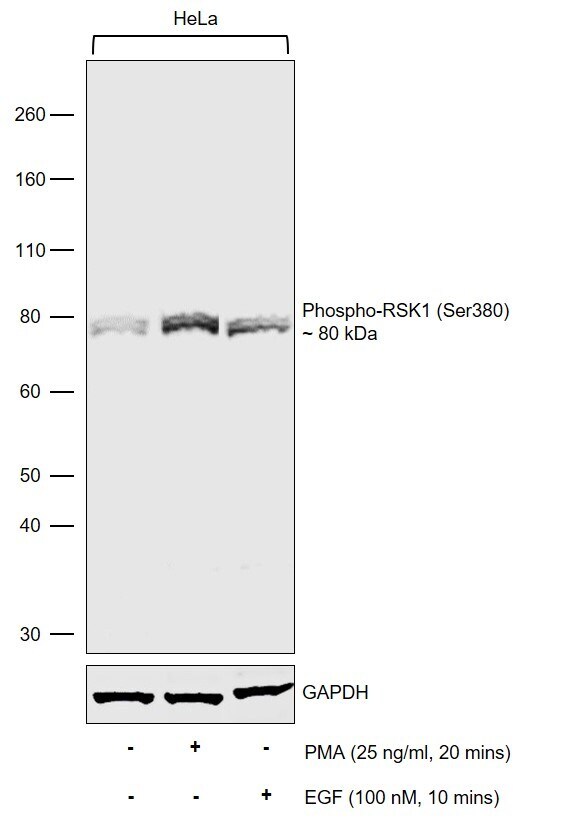
- Submitted by
- Invitrogen Antibodies (provider)
- Main image
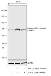
- Experimental details
- Western Blot was performed using Anti-Phospho-RSK1 (Ser380) Recombinant Rabbit Monoclonal Antibody (13H23L1) (Product # 702266) and a 80 kDa band corresponding to Phosphorylated Ribosomal protein S6 kinase alpha-1 was observed. Nuclear enriched extracts (30 µg lysate) of HeLa (Lane 1), HeLa treated with PMA (Over-night serum starvation followed by treatment with 25 ng/mL PMA for 20 mins) (Lane 2) and HeLa treated with EGF (Over-night serum starvation followed by treatment with 100 nM EGF for 10 mins) (Lane 3) were electrophoresed using NuPAGE™ 4-12% Bis-Tris Protein Gel (Product # NP0322BOX). Resolved proteins were then transferred onto a Nitrocellulose membrane (Product # IB23001) by iBlot® 2 Dry Blotting System (Product # IB21001). The Blot was probed with the primary antibody (1 µg/mL) and detected by chemiluminescence with Goat anti-Rabbit IgG (H+L) Superclonal™ Recombinant Secondary Antibody, HRP (Product # A27036, 1:4000) using the iBright FL 1000 (Product # A32752). Chemiluminescent detection was performed using Novex® ECL Chemiluminescent Substrate Reagent Kit (Product # WP20005). Up-regulation in the expression of phosphorylated RSK1 was observed in the lanes treated with PMA and EGF as expected (https://doi.org/10.1038/onc.2012.472, doi: 10.1091/mbc.E11-07-0658).