Explore
 Validate
 Learn
Learn33-1300
antibody from Invitrogen Antibodies
Targeting: MAPK14
CSBP1, CSBP2, CSPB1, Mxi2, p38, PRKM14, PRKM15
 Western blot
Antibody data
- Antibody Data
- Antigen structure
- References [9]
- Comments [0]
- Validations
- Western blot [2]
- Immunocytochemistry [1]
- Other assay [3]
Submit
Validation data
Reference
Comment
Report error
- Product number
- 33-1300 - Provider product page

- Provider
- Invitrogen Antibodies
- Product name
- p38 MAPK Monoclonal Antibody (p38-3F11)
- Antibody type
- Monoclonal
- Antigen
- Recombinant full-length protein
- Description
- 33-1300 has been successfully used in immunofluorescence, and western blot with human recombinant proteins and whole cell lysates of hamster, mouse, and primate origins.
- Antibody clone number
- p38-3F11
- Concentration
- 0.5 mg/mL
Submitted references Inhibiting Protein Kinase D Promotes Airway Epithelial Barrier Integrity in Mouse Models of Influenza A Virus Infection.
TGFβR-SMAD3 Signaling Induces Resistance to PARP Inhibitors in the Bone Marrow Microenvironment.
SkQ1 Suppresses the p38 MAPK Signaling Pathway Involved in Alzheimer's Disease-Like Pathology in OXYS Rats.
Wnt/β-catenin signaling pathway regulates asthma airway remodeling by influencing the expression of c-Myc and cyclin D1 via the p38 MAPK-dependent pathway.
Discovery of a Small Molecule Promoting Mouse and Human Osteoblast Differentiation via Activation of p38 MAPK-β.
Simulated Microgravity Disrupts Cytoskeleton Organization and Increases Apoptosis of Rat Neural Crest Stem Cells Via Upregulating CXCR4 Expression and RhoA-ROCK1-p38 MAPK-p53 Signaling.
Translational control of inducible nitric oxide synthase by p38 MAPK in islet β-cells.
Stress kinase phosphorylation is increased in pacing-induced heart failure in rabbits.
p38 mitogen-activated protein kinase is activated and linked to TNF-alpha signaling in inflammatory bowel disease.
Veazey JM, Eliseeva SI, Hillman SE, Stiles K, Smyth TR, Morrissey CE, Tillotson EJ, Topham DJ, Chapman TJ, Georas SN
Frontiers in immunology 2020;11:580401
Frontiers in immunology 2020;11:580401
TGFβR-SMAD3 Signaling Induces Resistance to PARP Inhibitors in the Bone Marrow Microenvironment.
Le BV, Podszywalow-Bartnicka P, Maifrede S, Sullivan-Reed K, Nieborowska-Skorska M, Golovine K, Yao JC, Nejati R, Cai KQ, Caruso LB, Swatler J, Dabrowski M, Lian Z, Valent P, Paietta EM, Levine RL, Fernandez HF, Tallman MS, Litzow MR, Huang J, Challen GA, Link D, Tempera I, Wasik MA, Piwocka K, Skorski T
Cell reports 2020 Oct 6;33(1):108221
Cell reports 2020 Oct 6;33(1):108221
SkQ1 Suppresses the p38 MAPK Signaling Pathway Involved in Alzheimer's Disease-Like Pathology in OXYS Rats.
Muraleva NA, Stefanova NA, Kolosova NG
Antioxidants (Basel, Switzerland) 2020 Jul 28;9(8)
Antioxidants (Basel, Switzerland) 2020 Jul 28;9(8)
Wnt/β-catenin signaling pathway regulates asthma airway remodeling by influencing the expression of c-Myc and cyclin D1 via the p38 MAPK-dependent pathway.
Jia XX, Zhu TT, Huang Y, Zeng XX, Zhang H, Zhang WX
Experimental and therapeutic medicine 2019 Nov;18(5):3431-3438
Experimental and therapeutic medicine 2019 Nov;18(5):3431-3438
Discovery of a Small Molecule Promoting Mouse and Human Osteoblast Differentiation via Activation of p38 MAPK-β.
Cook B, Rafiq R, Lee H, Banks KM, El-Debs M, Chiaravalli J, Glickman JF, Das BC, Chen S, Evans T
Cell chemical biology 2019 Jul 18;26(7):926-935.e6
Cell chemical biology 2019 Jul 18;26(7):926-935.e6
Simulated Microgravity Disrupts Cytoskeleton Organization and Increases Apoptosis of Rat Neural Crest Stem Cells Via Upregulating CXCR4 Expression and RhoA-ROCK1-p38 MAPK-p53 Signaling.
Lin SC, Gou GH, Hsia CW, Ho CW, Huang KL, Wu YF, Lee SY, Chen YH
Stem cells and development 2016 Aug 1;25(15):1172-93
Stem cells and development 2016 Aug 1;25(15):1172-93
Translational control of inducible nitric oxide synthase by p38 MAPK in islet β-cells.
Nishiki Y, Adewola A, Hatanaka M, Templin AT, Maier B, Mirmira RG
Molecular endocrinology (Baltimore, Md.) 2013 Feb;27(2):336-49
Molecular endocrinology (Baltimore, Md.) 2013 Feb;27(2):336-49
Stress kinase phosphorylation is increased in pacing-induced heart failure in rabbits.
Schulz R, Aker S, Belosjorow S, Konietzka I, Rauen U, Heusch G
American journal of physiology. Heart and circulatory physiology 2003 Nov;285(5):H2084-90
American journal of physiology. Heart and circulatory physiology 2003 Nov;285(5):H2084-90
p38 mitogen-activated protein kinase is activated and linked to TNF-alpha signaling in inflammatory bowel disease.
Waetzig GH, Seegert D, Rosenstiel P, Nikolaus S, Schreiber S
Journal of immunology (Baltimore, Md. : 1950) 2002 May 15;168(10):5342-51
Journal of immunology (Baltimore, Md. : 1950) 2002 May 15;168(10):5342-51
No comments: Submit comment
Supportive validation
- Submitted by
- Invitrogen Antibodies (provider)
- Main image

- Experimental details
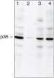
- Western blot analysis using Ms anti-p38a (Product # 33-1300) in: Lane 1: BHK cell lysates. Lane 2: C2C12 cell lysates. Lane 3: CHO cell lysates. Lane 4: COS cell lysates.
- Submitted by
- Invitrogen Antibodies (provider)
- Main image

- Experimental details
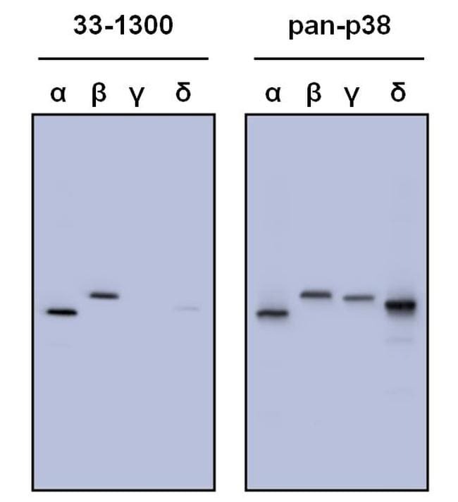
- Western blot analysis of p38 was performed by loading 50 ng of human, recombinant p38-alpha, beta, gamma and delta per well onto a Novex 4-20% Tris-Glycine polyacrylamide gel (Product # WT4202BOX ). Proteins were transferred to a nitrocellulose membrane using the G2 Blotter (Product # 62288), and blocked with 5% milk in TBST for one hour at room temperature. Isoforms of p38 were detected using a p38 monoclonal antibody (Product # 33-1300, left panel) and a pan-p38 antibody (right panel) at a dilution of 1 µg/mL in blocking buffer overnight at 4C on a rocking platform, followed by a Goat anti-mouse IgG HRP-linked secondary antibody (Product # 31430) at a dilution of 1:10,000 for at least 30 minutes. Chemiluminescent detection was performed using SuperSignal Pico (Product # 34078) and the myECL™ Imager (Product # 62236)
Supportive validation
- Submitted by
- Invitrogen Antibodies (provider)
- Main image
- Experimental details
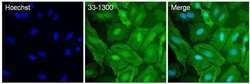
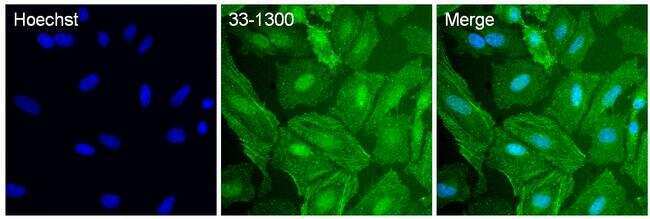
- Immunofluorescent analysis of p38-alpha (green) in HeLa cells. The cells were fixed with 4% paraformaldehyde for 15 minutes, permeabilized with 0.1% Triton X-100 in PBS for 10 minutes, and blocked with 3% BSA in PBS (Product # 37525) for 30 minutes at room temperature. Cells were stained with a mouse p38 monoclonal antibody (Product # 33-1300) at a dilution of 20 µg/mL in staining buffer for 1 hour at room temperature, and then incubated with a Goat anti-Mouse IgG (H+L) Superclonal Secondary Antibody, Alexa Fluor 488 conjugate (Product # A27027) at a dilution of 1:1000 for 1 hour at room temperature (green). Nuclei (blue) were stained with Hoechst 33342 dye (Product # 62249). Images were taken on a Thermo Scientific ToxInsight Instrument at 20X magnification.
Supportive validation
- Submitted by
- Invitrogen Antibodies (provider)
- Main image
- Experimental details
- NULL

- Submitted by
- Invitrogen Antibodies (provider)
- Main image
- Experimental details
- NULL

- Submitted by
- Invitrogen Antibodies (provider)
- Main image

- Experimental details
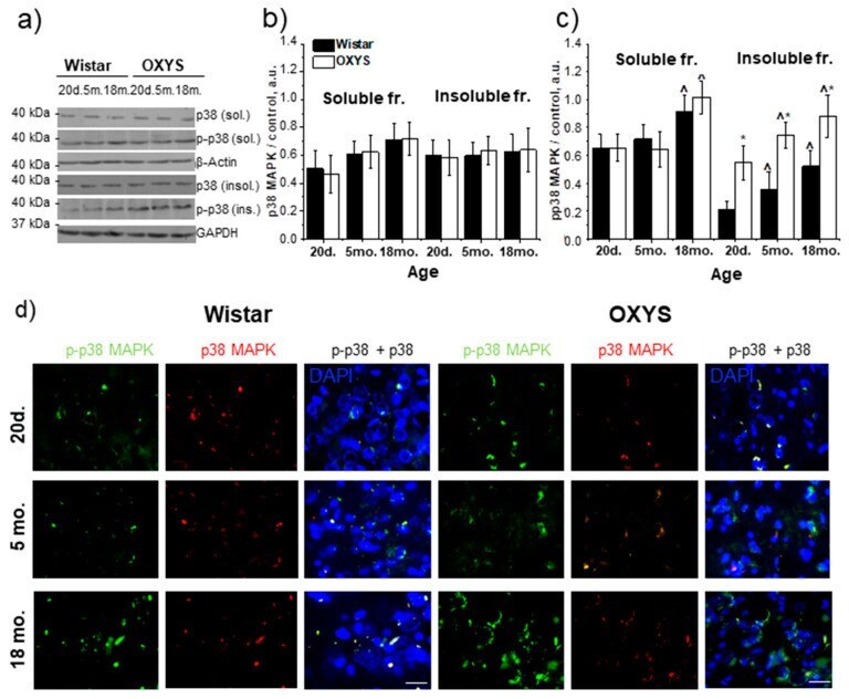
- Figure 2 The protein content of p38 MAPK and p-p38 MAPK in the hippocampus of 20-day-old and 5- and 18-month-old Wistar and OXYS rats. ( a ) Representative western blots of total and phosphorylated p38 MAPK in the detergent-soluble and detergent-insoluble fractions from the hippocampus of Wistar and OXYS rats. Graphical presentation illustrates the relative protein content of p38 MAPK ( b ) and p-p38 MAPK ( c ) in Wistar and OXYS rats' hippocampi at different ages after normalization of the detergent-soluble fraction data to beta-actin and detergent-insoluble fraction data to GAPDH. Data are presented as mean +- SEM of five independent experiments. Immunostaining for p38 MAPK and p-p38 MAPK ( d ) in the hippocampus of 20-day-old and 5- and 18-month-old Wistar and OXYS rats. The nuclei were stained with DAPI (blue). Scale bars, 25 um. * Statistically significant differences between the strains of the same age; ^ significant differences from the previous age within a strain ( p < 0.05).