Explore
 Validate
 Learn
Learn Western blot
Antibody data
- Antibody Data
- Antigen structure
- References [5]
- Comments [0]
- Validations
- Western blot [1]
- Immunocytochemistry [2]
- Immunohistochemistry [1]
- Other assay [2]
Submit
Validation data
Reference
Comment
Report error
- Product number
- PA1-16751 - Provider product page

- Provider
- Invitrogen Antibodies
- Product name
- MAP2 Polyclonal Antibody
- Antibody type
- Polyclonal
- Antigen
- Recombinant full-length protein
- Description
- In Western blot bands can be seen at approximately 280 kDa, representing MAP2a and MAP2b.
Submitted references Capping Protein Regulator and Myosin 1 Linker 3 (CARMIL3) as a Molecular Signature of Ischemic Neurons in the DWI-T2 Mismatch Areas After Stroke.
Pervasive compartment-specific regulation of gene expression during homeostatic synaptic scaling.
Comparison of induced neurons reveals slower structural and functional maturation in humans than in apes.
Structure-Activity Relationship of Neuroactive Steroids, Midazolam, and Perampanel Toward Mitigating Tetramine-Triggered Activity in Murine Hippocampal Neuronal Networks.
Human Cerebrospinal Fluid Monoclonal LGI1 Autoantibodies Increase Neuronal Excitability.
Yeh SJ, Hsu PH, Yeh TY, Yang WK, Chang KP, Chiang CS, Tang SC, Tsai LK, Jeng JS, Hsieh ST
Frontiers in molecular neuroscience 2021;14:754762
Frontiers in molecular neuroscience 2021;14:754762
Pervasive compartment-specific regulation of gene expression during homeostatic synaptic scaling.
Colameo D, Rajman M, Soutschek M, Bicker S, von Ziegler L, Bohacek J, Winterer J, Germain PL, Dieterich C, Schratt G
EMBO reports 2021 Oct 5;22(10):e52094
EMBO reports 2021 Oct 5;22(10):e52094
Comparison of induced neurons reveals slower structural and functional maturation in humans than in apes.
Schörnig M, Ju X, Fast L, Ebert S, Weigert A, Kanton S, Schaffer T, Nadif Kasri N, Treutlein B, Peter BM, Hevers W, Taverna E
eLife 2021 Jan 20;10
eLife 2021 Jan 20;10
Structure-Activity Relationship of Neuroactive Steroids, Midazolam, and Perampanel Toward Mitigating Tetramine-Triggered Activity in Murine Hippocampal Neuronal Networks.
Antrobus S, Pressly B, Nik AM, Wulff H, Pessah IN
Toxicological sciences : an official journal of the Society of Toxicology 2021 Apr 12;180(2):325-341
Toxicological sciences : an official journal of the Society of Toxicology 2021 Apr 12;180(2):325-341
Human Cerebrospinal Fluid Monoclonal LGI1 Autoantibodies Increase Neuronal Excitability.
Kornau HC, Kreye J, Stumpf A, Fukata Y, Parthier D, Sammons RP, Imbrosci B, Kurpjuweit S, Kowski AB, Fukata M, Prüss H, Schmitz D
Annals of neurology 2020 Mar;87(3):405-418
Annals of neurology 2020 Mar;87(3):405-418
No comments: Submit comment
Supportive validation
- Submitted by
- Invitrogen Antibodies (provider)
- Main image
- Experimental details
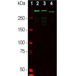
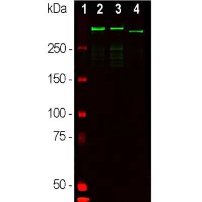
- Western blot analysis of MAP2 in whole brain tissue lysates. Samples were incubated in MAP2 polyclonal antibody (Product # PA1-16751 using a dilution of 1:50,000. Antibody in green: [1] protein standard (red), [2] adult rat brain, [3] embryonic E20 rat brain, [4] adult mouse brain. Strong band observed at ~280 kDa mark corresponds to two major isoforms of MAP2 protein referred to as MAP2A and MAP2B. Smaller fragments of these isoforms are also detected if the antibody is used at higher concentrations.
Supportive validation
- Submitted by
- Invitrogen Antibodies (provider)
- Main image

- Experimental details
- Immunocytochemistry analysis of MAP2 in rat hippocampi. Samples were incubated in MAP2 polyclonal antibody (Product # PA1-16751). PEA decreases astrocyte activation in organotypic cultures of rat hippocampi and rescues neuronal CA3 damage caused by Abeta challenge. Abeta-challenged (1 µg/mL) slices of rat hippocampi were treated for 24 hours with PEA (0.1 µM) in the presence of the selective PPAR antagonist (GW9662, 9 nM) or the selective PPAR alpha antagonist (MK886, 3 µM). Representative photomicrographs of the CA3 region showing the results of immunofluorescence experiments aimed at investigating the effect of treatments on astrocyte activation and neuronal loss, as determined by immunostaining for GFAP (green) and MAP2 (red) alone or merged, respectively. Nuclei were stained with Hoechst (blue). Scale bar: 10 µm. Arrows in the photomicrographs indicate astrocyte infiltration events and apoptotic condensation in the nuclei of adjacent neurons.

- Submitted by
- Invitrogen Antibodies (provider)
- Main image
- Experimental details
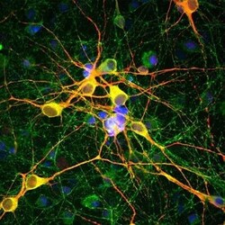
- Immunocytochemistry analysis of MAP2 in cortical neuron-glial cell culture from E20 rat. Samples were incubated in MAP2 polyclonal antibody (Product # PA1-16751) using a dilution of 1:10000. This antibody in red, and mouse mAb to MAP-tau dilution 1:2,000, in green. The blue is DAPI staining of nuclear DNA. This antibody stains dendrites and perikarya of neurons, while Tau antibody labels neuronal perikarya, dendrites and also axonal process. As a result perikarya and dendrites appears orange-yellow, since they contain both MAP2 and tau, while axons are green.

Supportive validation
- Submitted by
- Invitrogen Antibodies (provider)
- Main image

- Experimental details
- View of mixed neuron/glial cultures stained with CPCA-MAP2 (red). The perikarya and dendrites of neurons are strongly and specifically stained with the MAP2 antibody, while the axons of the neurons and the processes of all other cell types in these cultures (astrocytes, oligodendrocytes, microglia, endothelia and fibroblasts) are all negative. Cell nuclei are visualized with DAPI DNA stain.

Supportive validation
- Submitted by
- Invitrogen Antibodies (provider)
- Main image
- Experimental details
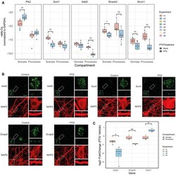
- Figure 3 Validation of compartment-specific regulations Real-time quantitative PCR (RT-qPCR) of transcripts changing differentially between compartments using the compartmentalized cultures after 48 h PTX treatment. n = 8 independent biological replicates (except for Plk2 ( n = 7), where one measurement couldn' t be performed due to insufficient cDNA); PTX effect was assessed by three-way ANOVA followed by Tukey's post hoc multiple comparison test; * P < 0.05; ** P < 0.01. Representative images of single-molecule FISH (smFISH) in either control or 48 h PTX-treated rat hippocampal neurons (DIV20) using probes specific for Add2, Dnajc6 and Sort1 (green). MAP2 immunostaining (red) was used to visualize neuronal somata and dendrites. Inserts at higher magnification illustrate PTX-dependent changes in dendritic RNA puncta. Scale bar = 10 mum. Quantification of (B). ( n = 3-4 independent biological replicates with 8-10 cells averaged per condition and replicate; two-sample Student's t -test; * P < 0.05; ** P < 0.01). Data information: Boxplots: central line: median; box: 25 th to 75 th percentile; whiskers: until last data point within 1.5x interquartile range (IQR).

- Submitted by
- Invitrogen Antibodies (provider)
- Main image

- Experimental details
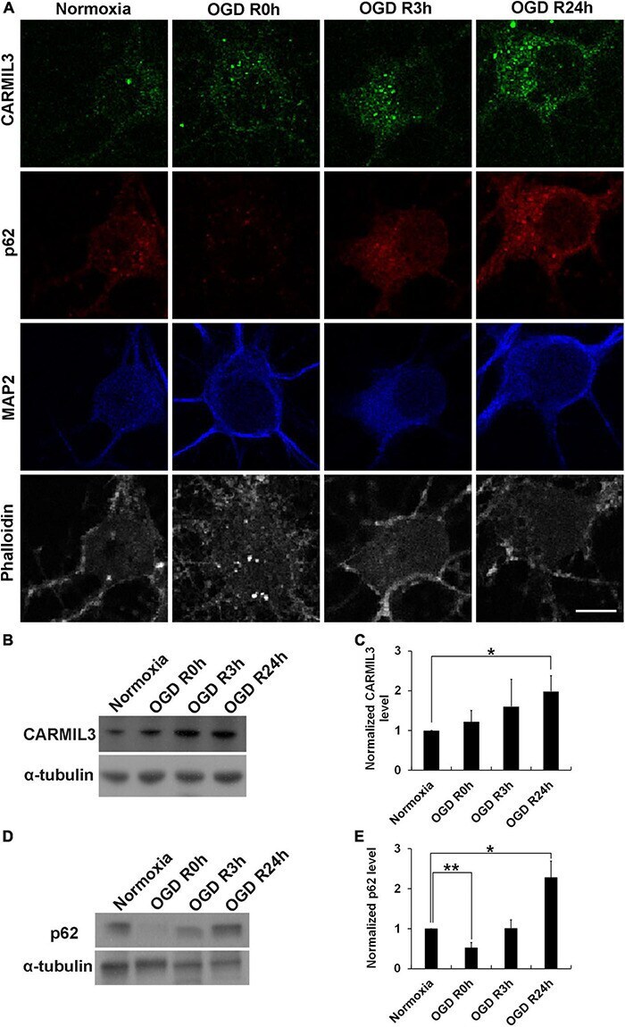
- FIGURE 11 CARMIL3 expression in rat primary cortical neuronal culture. (A) Immunofluorescence staining of CARMIL3, p62, MAP2, and phalloidin in primary rat cortical neuron culture after oxygen-glucose deprivation (OGD). CARMIL3 was obviously expressed as a beaded pattern in the neurons at OGD R3h and OGD R24h; p62 had increased expression in neurons at the two points in time. There was more phalloidin staining at OGD R3h, indicating more expression of filamentous actin. (B,C) Western blot for CARMIL3 in neuronal culture showed that OGD R24h had significantly increased CARMIL3 levels than the normoxia group (normalized expression level 1.98 vs. 1, p = 0.025, n = 3; p = 0.065 in comparison among the 4 groups). (D,E) Western blot for p62 in neuronal culture revealed lower expression in the OGD R0h group while higher expression in the OGD R24h group compared with that in the normoxia group. * p < 0.05; ** p < 0.01. Scale bar = 10 mum.