 Explore
 Validate
 Learn
Learn Western blot
Antibody data
- Antibody Data
- Antigen structure
- References [0]
- Comments [0]
- Validations
- Western blot [5]
- Immunocytochemistry [2]
- Immunohistochemistry [1]
Submit
Validation data
Reference
Comment
Report error
- Product number
- PA5-30183 - Provider product page

- Provider
- Invitrogen Antibodies
- Product name
- MUNC18 Polyclonal Antibody
- Antibody type
- Polyclonal
- Antigen
- Recombinant protein fragment
- Description
- Recommended positive controls: A431, mouse brain, rat brain.
- Concentration
- 0.8 mg/mL
No comments: Submit comment
Supportive validation
- Submitted by
- Invitrogen Antibodies (provider)
- Main image
- Experimental details
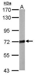
- Western blot analysis of STXBP1 using 20 µg of mouse brain lysate. Samples were loaded onto a 7.5% SDS-PAGE gel and probed with a STXBP1 polyclonal antibody (Product # PA5-30183) at a dilution of 1:10,000.

- Submitted by
- Invitrogen Antibodies (provider)
- Main image
- Experimental details
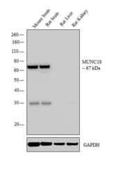
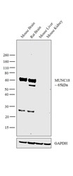
- Western blot analysis was performed on whole cell extract (30 µg lysate) of Mouse brain (Lane 1), Rat brain (Lane 2), Rat Liver (Lane 3) and Rat Kidney (Lane 4). The blot was probed with Anti-MUNC18 Polyclonal Antibody (Product # PA5-30183, 1:5000 dilution) and detected by chemiluminescence using Goat anti-Rabbit IgG (H+L) Superclonal™ Secondary Antibody, HRP conjugate (Product # A27036, 0.25 µg/mL, 1:4000 dilution). A 67 kDa band corresponding to MUNC18 was observed in the Mouse and Rat brain, while it was not detected in Rat Liver and Kidney which are reported negative for MUNC18 expression. An additional band was observed at ~28kDa in mouse and Rat brain tissues.

- Submitted by
- Invitrogen Antibodies (provider)
- Main image
- Experimental details
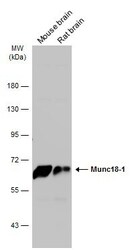
- Western Blot analysis of MUNC18 was performed by separating 50 µg of Various tissue extracts by 7.5% SDS-PAGE. Proteins were transferred to a membrane and probed with a MUNC18 Polyclonal Antibody (Product # PA5-30183) at a dilution of 1:10000. The HRP-conjugated anti-rabbit IgG antibody was used to detect the primary antibody.
- Submitted by
- Invitrogen Antibodies (provider)
- Main image

- Experimental details
- Western Blot using MUNC18 Polyclonal Antibody (Product # PA5-30183). Sample (30 µg of whole cell lysate). Lane A: A431 . 7.5% SDS PAGE. MUNC18 Polyclonal Antibody (Product # PA5-30183) diluted at 1:1,000.

- Submitted by
- Invitrogen Antibodies (provider)
- Main image
- Experimental details
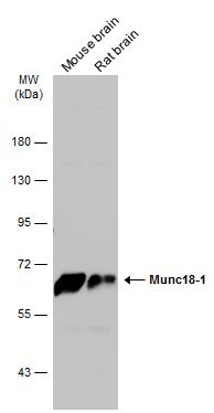
- Western blot analysis was performed on whole cell extracts (30 µg lysate) of Mouse Brain (Lane 1), Rat Brain (Lane 2), Mouse Liver (Lane 3) and Mouse Kidney (Lane 4). The blot was probed with Anti-MUNC18 Polyclonal Antibody (Product # PA5-30183, 1:5000 dilution) and detected by chemiluminescence using Goat anti-Rabbit IgG (H+L) Superclonal™ Secondary Antibody, HRP conjugate (Product # A27036, 0.25 µg/mL, 1:4000 dilution). A 65kDa band corresponding to MUNC18 was seen in all tissue lysates tested except Mouse Liver and Mouse Kidney which are reported to be negative for MUNC18 expression. An additional band as also observed at ~28kDa in brain samples.

Supportive validation
- Submitted by
- Invitrogen Antibodies (provider)
- Main image

- Experimental details
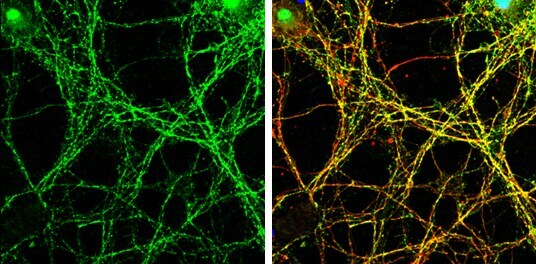
- Immunocytochemistry-Immunofluorescence analysis of MUNC18 was performed in DIV9 rat E18 primary cortical neurons fixed in 4% paraformaldehyde at RT for 15 min. Green: MUNC18 Polyclonal Antibody (Product # PA5-30183) diluted at 1:500. Red: beta Tubulin 3/ Tuj1, stained by beta Tubulin 3/ Tuj1 antibody. Blue: Fluoroshield with DAPI.
- Submitted by
- Invitrogen Antibodies (provider)
- Main image
- Experimental details
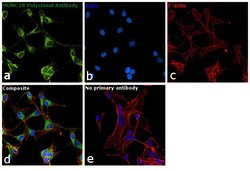
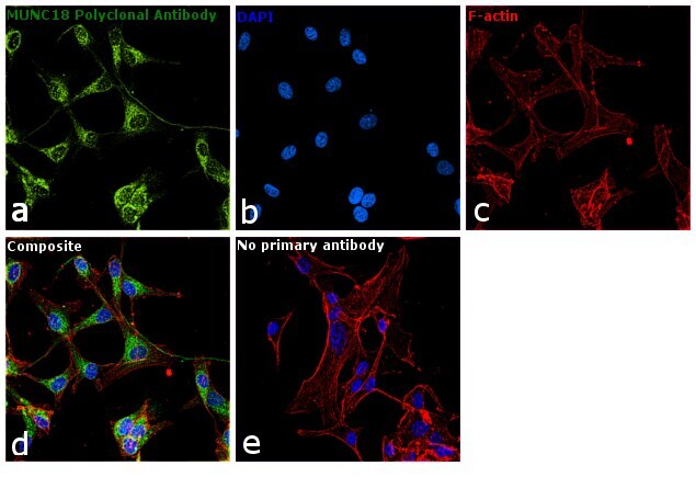
- Immunofluorescence analysis of MUNC18 was performed using 70% confluent log phase U-87 MG cells. The cells were fixed with 4% paraformaldehyde for 10 minutes, permeabilized with 0.1% Triton™ X-100 for 15 minutes, and blocked with 1% BSA for 1 hour at room temperature. The cells were labeled with MUNC18 Polyclonal Antibody (Product # PA5-30183) at 1:100 dilution in 0.1% BSA, incubated at 4 degree Celsius overnight and then labeled with Goat anti-Rabbit IgG (H+L) Superclonal™ Secondary Antibody, Alexa Fluor® 488 conjugate (Product # A27034) at a dilution of 1:2000 for 45 minutes at room temperature (Panel a: green). Nuclei (Panel b: blue) were stained with ProLong™ Diamond Antifade Mountant with DAPI (Product # P36962). F-actin (Panel c: red) was stained with Rhodamine Phalloidin (Product # R415). Panel d represents the merged image showing Cytoplasmic and Nuclear localization. Panel e represents control cells with no primary antibody to assess background. The images were captured at 60X magnification.
Supportive validation
- Submitted by
- Invitrogen Antibodies (provider)
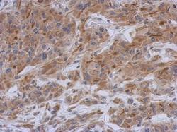
- Main image
- Experimental details
- Immunohistochemical analysis of paraffin-embedded U373 xenograft, using STXBP1 (Product # PA5-30183) antibody at 1:500 dilution. Antigen Retrieval: EDTA based buffer, pH 8.0, 15 min.
