Explore
 Validate
 Learn
Learn Western blot
Antibody data
- Antibody Data
- Antigen structure
- References [0]
- Comments [0]
- Validations
- Western blot [7]
- Immunocytochemistry [1]
- Immunohistochemistry [2]
Submit
Validation data
Reference
Comment
Report error
- Product number
- PA5-27323 - Provider product page

- Provider
- Invitrogen Antibodies
- Product name
- VCP Polyclonal Antibody
- Antibody type
- Polyclonal
- Antigen
- Recombinant protein fragment
- Description
- Recommended positive controls: A431, H1299, mouse brain, rat brain, A549, H1299, HCT-116.
- Concentration
- 1 mg/mL
No comments: Submit comment
Supportive validation
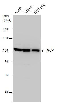
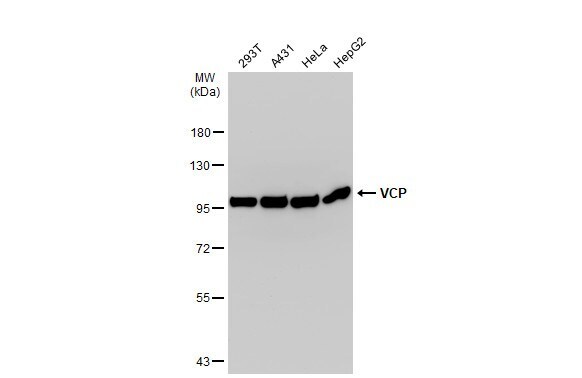
- Submitted by
- Invitrogen Antibodies (provider)
- Main image
- Experimental details
- Western blot analysis of VCP using Various whole cell extracts (30 µg). Samples were loaded onto a 7.5% SDS-PAGE gel and probed with a VCP polyclonal antibody (Product # PA5-27323) at a dilution of 1:2000.
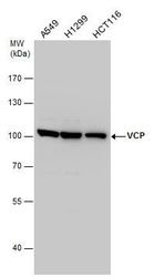
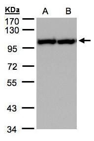
- Submitted by
- Invitrogen Antibodies (provider)
- Main image
- Experimental details
- Western blot analysis of VCP using 30 µg of A) A431 and B) H1299 lysate. Samples were loaded onto a 7.5% SDS-PAGE gel and probed with a VCP polyclonal antibody (Product # PA5-27323) at a dilution of 1:2000.
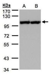
- Submitted by
- Invitrogen Antibodies (provider)
- Main image
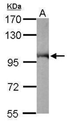
- Experimental details
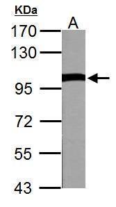
- Western Blot using VCP Polyclonal Antibody (Product # PA5-27323). Sample (50 µg of whole cell lysate). Lane A: Mouse brain. 7.5% SDS PAGE. VCP Polyclonal Antibody (Product # PA5-27323) diluted at 1:10,000. The HRP-conjugated anti-rabbit IgG antibody was used to detect the primary antibody.

- Submitted by
- Invitrogen Antibodies (provider)
- Main image
- Experimental details
- Western Blot using VCP Polyclonal Antibody (Product # PA5-27323). Various whole cell extracts (30 µg) were separated by 7.5% SDS-PAGE, and the membrane was blotted with VCP Polyclonal Antibody (Product # PA5-27323) diluted at 1:500. The HRP-conjugated anti-rabbit IgG antibody was used to detect the primary antibody.
- Submitted by
- Invitrogen Antibodies (provider)
- Main image
- Experimental details
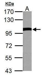
- VCP Polyclonal Antibody detects VCP protein by western blot analysis. A. 50 µg Rat brain lysate/extract.7.5% SDS-PAGE. VCP Polyclonal Antibody (Product # PA5-27323) dilution: 1:10,000. The HRP-conjugated anti-rabbit IgG antibody was used to detect the primary antibody.
- Submitted by
- Invitrogen Antibodies (provider)
- Main image
- Experimental details
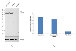
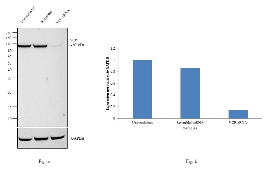
- Knockdown of VCP was achieved by transfecting MCF7 cells with VCP specific siRNAs (Silencer® select Product # s14765). Western blot analysis (Fig. a) was performed using whole cell extracts from the VCP knockdown cells (lane 3), non-specific scrambled siRNA transfected cells (lane 2) and untransfected cells (lane 1). The blot was probed with VCP Polyclonal Antibody (Product # PA5-27323, 1:8000 dilution) and Goat anti-Rabbit IgG (H+L) Superclonal™ Secondary Antibody, HRP conjugate (Product # A27036, 0.25µg/ml, 1:4000 dilution). Densitometric analysis of this western blot is shown in histogram (Fig. b). Decrease in signal upon siRNA mediated knock down confirms that antibody is specific to VCP.
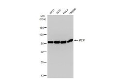
- Submitted by
- Invitrogen Antibodies (provider)
- Main image
- Experimental details
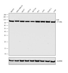
- Western blot analysis was performed on whole cell extract (30 µg lysate) of SKOV3 (Lane 1), MDA-MB-231 (Lane 2), MCF7 (Lane 3), GTL16 (Lane 4), HCT116 (Lane 5), PC3 (Lane 6), A431 (Lane 7), PANC-1 (Lane 8) and A549 (Lane 9). The blot was probed with Anti-VCP Polyclonal Antibody (Product # PA5-27323, 1:8000 dilution) and detected by chemiluminescence using Goat anti-Rabbit IgG (H+L) Superclonal™ Secondary Antibody, HRP conjugate (Product # A27036, 0.25 µg/ml, 1:4000 dilution). A 97 kDa band corresponding to VCP was observed in all cell lines.
Supportive validation
- Submitted by
- Invitrogen Antibodies (provider)
- Main image

- Experimental details
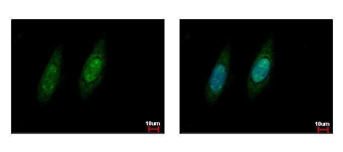
- VCP Polyclonal Antibody detects VCP protein at cytoplasm and nucleus by immunofluorescent analysis. Sample: HeLa cells were fixed in ice-cold MeOH for 5 min. Green: VCP protein stained by VCP Polyclonal Antibody (Product # PA5-27323) diluted at 1:500. Blue: Hoechst 33343 staining.
Supportive validation
- Submitted by
- Invitrogen Antibodies (provider)
- Main image
- Experimental details
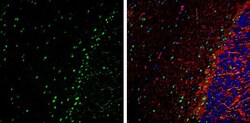
- Immunohistochemistry (Frozen) analysis of VCP was performed in frozen-sectioned adult mouse cerebellum tissue using VCP Polyclonal Antibody (Product # PA5-27323) at a dilution of 1:250 (Green). Red: NF-H, stained by NF-H antibody diluted at 1:500. Blue: Fluoroshield with DAPI. Antigen Retrieval: Citrate buffer, pH 6.0, 10 min.

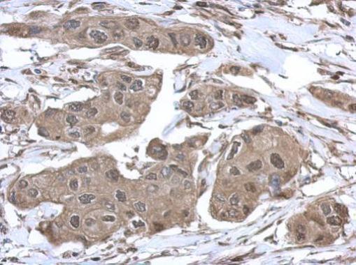
- Submitted by
- Invitrogen Antibodies (provider)
- Main image

- Experimental details
- Immunohistochemical analysis of paraffin-embedded human breast cancer, using VCP (Product # PA5-27323) antibody at 1:500 dilution. Antigen Retrieval: EDTA based buffer, pH 8.0, 15 min.