Explore
 Validate
 Learn
Learn Western blot
Antibody data
- Antibody Data
- Antigen structure
- References [0]
- Comments [0]
- Validations
- Western blot [3]
Submit
Validation data
Reference
Comment
Report error
- Product number
- PA5-39270 - Provider product page

- Provider
- Invitrogen Antibodies
- Product name
- Anti-NDUFA9 Polyclonal Antibody
- Antibody type
- Polyclonal
- Antigen
- Synthetic peptide
- Reactivity
- Human, Mouse, Rat
- Host
- Rabbit
- Isotype
- IgG
- Vial size
- 100 µg
- Concentration
- 1 mg/mL
- Storage
- -20°C
No comments: Submit comment
Supportive validation
- Submitted by
- Invitrogen Antibodies (provider)
- Main image
- Experimental details
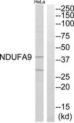
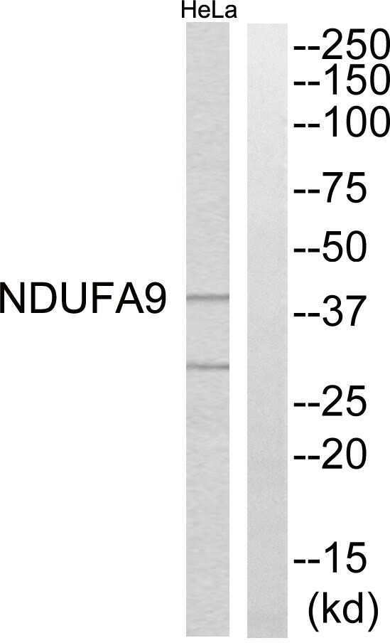
- Western blot analysis of NDUFA9 in extracts from HeLa cells using a NDUFA9 polyclonal antibody (Product # PA5-39270).
- Submitted by
- Invitrogen Antibodies (provider)
- Main image
- Experimental details
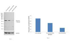
- KD of NDUFA9 was achieved by transfecting Hep G2 cells with NDUFA9 specific siRNAs (Silencer® select Product # s39346, s9347). Western blot analysis (Fig. a) was performed using membrane enriched cell extracts from the NDUFA9 KD cells (lane 3), non-specific scrambled siRNA transfected cells (lane 2) and untransfected cells (lane 1). The blot was probed NDUFA9 Polyclonal Antibody (Product # PA5-39270, 1:1000 dilution) and Goat anti-Rabbit IgG (H+L) Superclonal™ Secondary Antibody, HRP conjugate (Product # A27036, 0.25 µg/mL, 1:4000 dilution). Densitometric analysis of this western blot is shown in histogram (Fig. b). Decrease in signal upon siRNA mediated knock down confirms that antibody is specific to NDUFA9.

- Submitted by
- Invitrogen Antibodies (provider)
- Main image
- Experimental details
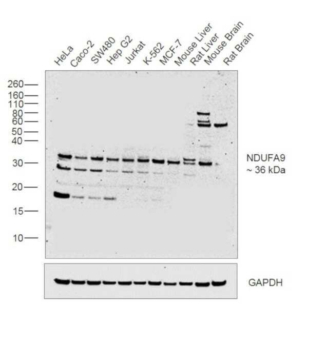
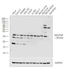
- Western blot was performed using Anti-NDUFA9 Polyclonal Antibody (Product # PA5-39270) and a 36 kDa band corresponding to NDUFA9 was observed across cell lines and tissues tested. Membrane enriched cell extracts (30 µg lysate) of HeLa (Lane 1), Caco-2 (Lane 2), SW480 (Lane 3), Hep G2 (Lane 4), Jurkat (Lane 5), K-562 (Lane 6), MCF-7 (Lane 7), Mouse Liver (Lane 8), Rat Liver (Lane 9), Mouse Brain (Lane 10) and Rat Brain (Lane 11) were electrophoresed using Novex® NuPAGE® 12 % Bis-Tris gel (Product # NP0342BOX). Resolved proteins were then transferred onto a nitrocellulose membrane by iBlot® 2 Dry Blotting System (Product # IB21001). The blot was probed with the primary antibody (1:1000 dilution) and detected by chemiluminescence using Goat Anti-Rabbit IgG Secondary Antibody, HRP conjugate (Product # A27036, 1:4000 dilution) using the iBright FL 1000 (Product # A32752). Chemiluminescent detection was performed using Novex® ECL Chemiluminescent Substrate Reagent Kit (Product # WP20005).