Explore
 Validate
 Learn
Learn Western blot
Antibody data
- Antibody Data
- Antigen structure
- References [3]
- Comments [0]
- Validations
- Western blot [7]
- Immunohistochemistry [3]
- Other assay [4]
Submit
Validation data
Reference
Comment
Report error
- Product number
- PA5-22113 - Provider product page

- Provider
- Invitrogen Antibodies
- Product name
- CD74 Polyclonal Antibody
- Antibody type
- Polyclonal
- Antigen
- Synthetic peptide
- Description
- Recommended positive controls: Raji, GL261, Rat2.
- Concentration
- 0.87 mg/mL
Submitted references Commensal microbiota divergently affect myeloid subsets in the mammalian central nervous system during homeostasis and disease.
Immune-modulating Activity of Hydrogel Microparticles Contributes to the Host Defense in a Murine Model of Cutaneous Anthrax.
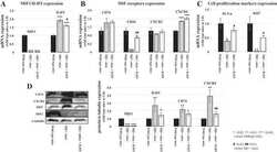
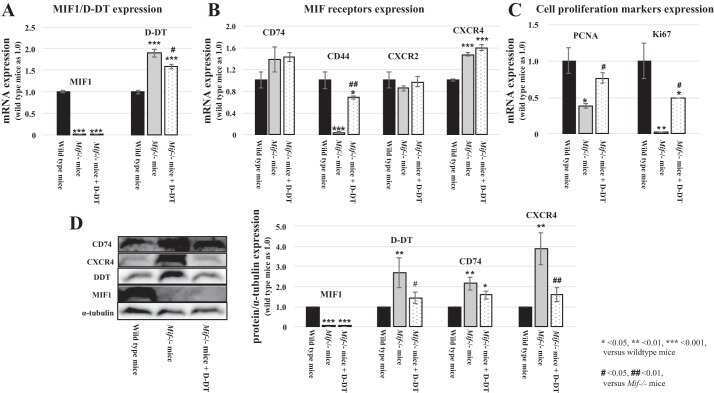
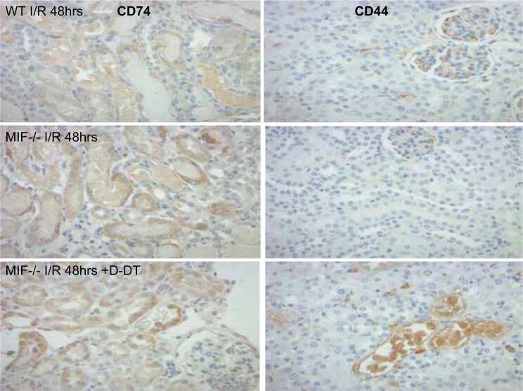
MIF-2/D-DT enhances proximal tubular cell regeneration through SLPI- and ATF4-dependent mechanisms.
Sankowski R, Ahmari J, Mezö C, Hrabě de Angelis AL, Fuchs V, Utermöhlen O, Buch T, Blank T, Gomez de Agüero M, Macpherson AJ, Erny D
The EMBO journal 2021 Dec 1;40(23):e108605
The EMBO journal 2021 Dec 1;40(23):e108605
Immune-modulating Activity of Hydrogel Microparticles Contributes to the Host Defense in a Murine Model of Cutaneous Anthrax.
Teunis AL, Popova TG, Espina V, Liotta LA, Popov SG
Frontiers in molecular biosciences 2017;4:62
Frontiers in molecular biosciences 2017;4:62
MIF-2/D-DT enhances proximal tubular cell regeneration through SLPI- and ATF4-dependent mechanisms.
Ochi A, Chen D, Schulte W, Leng L, Moeckel N, Piecychna M, Averdunk L, Stoppe C, Bucala R, Moeckel G
American journal of physiology. Renal physiology 2017 Sep 1;313(3):F767-F780
American journal of physiology. Renal physiology 2017 Sep 1;313(3):F767-F780
No comments: Submit comment
Supportive validation
- Submitted by
- Invitrogen Antibodies (provider)
- Main image
- Experimental details
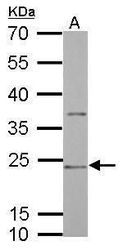
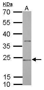
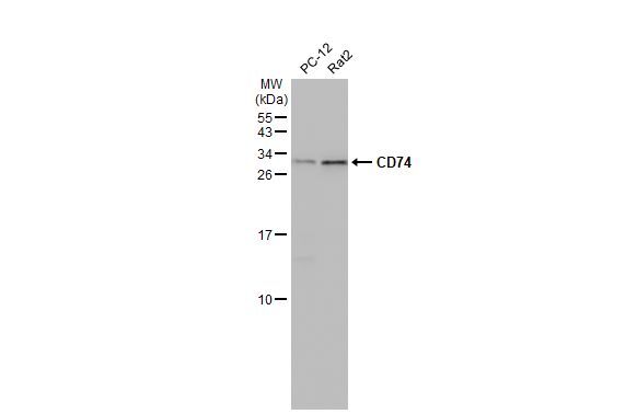
- Western blot analysis of MHC II HLA-DR/CD74 using 30 µg GL261 whole cell lysate. Samples were loaded onto a 12% SDS-PAGE gel and probed with a MHC II HLA-DR/CD74 polyclonal antibody (Product # PA5-22113) at a dilution of 1:1000.

- Submitted by
- Invitrogen Antibodies (provider)
- Main image
- Experimental details
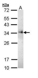
- Western blot analysis of MHC II HLA-DR/CD74 using 30 µg Rat2 whole cell lysate. Samples were loaded onto a 12% SDS-PAGE gel and probed with a MHC II HLA-DR/CD74 polyclonal antibody (Product # PA5-22113) at a dilution of 1:1000.
- Submitted by
- Invitrogen Antibodies (provider)
- Main image
- Experimental details
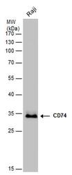
- Western blot analysis of MHC II HLA-DR/CD74 using 30 µg of Raji lysate. Samples were loaded onto a 12% SDS-PAGE gel and probed with a MHC II HLA-DR/CD74 polyclonal antibody (Product # PA5-22113) at a dilution of 1:5000.

- Submitted by
- Invitrogen Antibodies (provider)
- Main image
- Experimental details
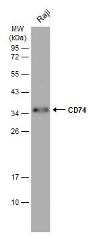
- Western blot analysis of CD74 in whole cell extracts (30 µg). Samples was separated by 12% SDS-PAGE and the membrane was probed with CD74 Polyclonal antibody (Product # PA5-22113) at a dilution of 1:5000.

- Submitted by
- Invitrogen Antibodies (provider)
- Main image

- Experimental details
- Western blot analysis of CD74 was performed by separating 30 µg of whole cell extract by 12% SDS-PAGE. Proteins were transferred to a membrane and probed with a CD74 Polyclonal Antibody (Product # PA5-22113) at a dilution of 1:1000. The HRP-conjugated anti-rabbit IgG antibody was used to detect the primary antibody.
- Submitted by
- Invitrogen Antibodies (provider)
- Main image

- Experimental details
- Western Blot using CD74 Polyclonal Antibody (Product # PA5-22113). Various whole cell extracts (30 µg) were separated by 15% SDS-PAGE, and the membrane was blotted with CD74 Polyclonal Antibody (Product # PA5-22113) diluted at 1:1,000. The HRP-conjugated anti-rabbit IgG antibody was used to detect the primary antibody.
- Submitted by
- Invitrogen Antibodies (provider)
- Main image
- Experimental details
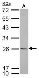
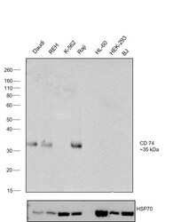
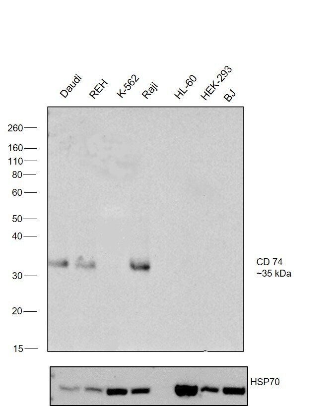
- Western blot was performed using Anti-CD74 Polyclonal Antibody, (Product # PA5-22113) and 35 kDa band corresponding to CD 74 was observed across cell lines tested except in K-562, HL-60, HEK-293 and BJ. Whole cell extracts (30 µg lysate) of Daudi (Lane 1), REH (Lane 2), K-562 (Lane 3), Raji (Lane 4), HL-60 (Lane 5), HEK-293 (Lane 6) and BJ (Lane 7) were electrophoresed using Novex® NuPAGE® 4-12 % Bis-Tris gel (Product # NP0321BOX). Resolved proteins were then transferred onto a nitrocellulose membrane (Product # IB23001) by iBlot® 2 Dry Blotting System (Product # IB21001). The blot was probed with the primary antibody (1.35µg/mL) and detected by chemiluminescence with Goat anti-Rabbit IgG (H+L), Superclonal™ Recombinant Secondary Antibody, HRP conjugate (Product # A27036, 1:4000 dilution) using the iBright FL 1000 (Product # A32752). Chemiluminescent detection was performed using Novex® ECL Chemiluminescent Substrate Reagent Kit (Product # WP20005).
Supportive validation
- Submitted by
- Invitrogen Antibodies (provider)
- Main image
- Experimental details
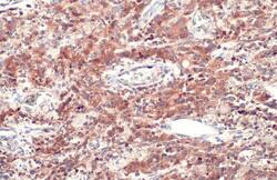
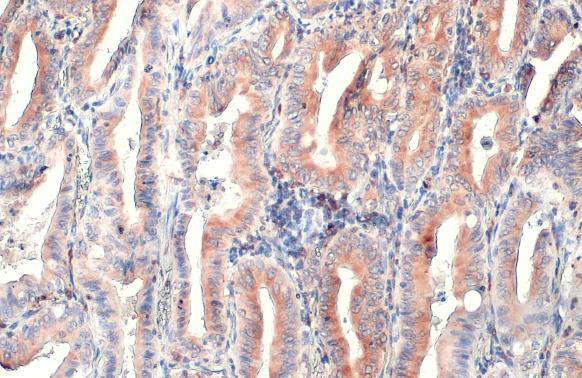
- CD74 Polyclonal Antibody detects CD74 protein at cytoplasm by immunohistochemical analysis. Sample: Paraffin-embedded human lung cancer. CD74 stained by CD74 Polyclonal Antibody (Product # PA5-22113) diluted at 1:500. Antigen Retrieval: Citrate buffer, pH 6.0, 15 min.

- Submitted by
- Invitrogen Antibodies (provider)
- Main image
- Experimental details
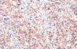
- CD74 Polyclonal Antibody detects CD74 protein at cytoplasm by immunohistochemical analysis. Sample: Paraffin-embedded human lung cancer. CD74 stained by CD74 Polyclonal Antibody (Product # PA5-22113) diluted at 1:500. Antigen Retrieval: Citrate buffer, pH 6.0, 15 min.

- Submitted by
- Invitrogen Antibodies (provider)
- Main image

- Experimental details
- CD74 Polyclonal Antibody detects CD74 protein at cytoplasm by immunohistochemical analysis. Sample: Paraffin-embedded human lung cancer. CD74 stained by CD74 Polyclonal Antibody (Product # PA5-22113) diluted at 1:500. Antigen Retrieval: Citrate buffer, pH 6.0, 15 min.
Supportive validation
- Submitted by
- Invitrogen Antibodies (provider)
- Main image
- Experimental details
- NULL
- Submitted by
- Invitrogen Antibodies (provider)
- Main image
- Experimental details
- NULL
- Submitted by
- Invitrogen Antibodies (provider)
- Main image
- Experimental details
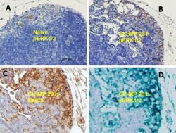
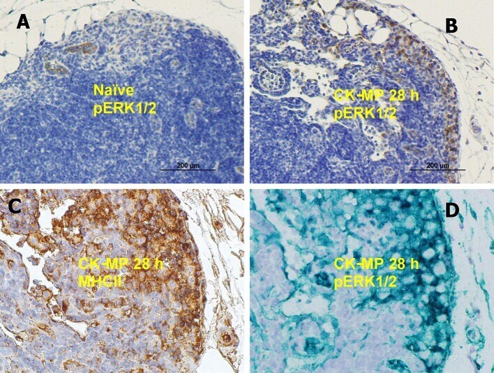
- Figure 5 (A,B) Medullary region of popliteal LN in naive mice (A) or after injection of CK-MPs for 28 h (B) 40x. Immunohistochemical staining with pERK1/2-specific antibody. (A,B,D) Overlapping patterns of immunohistochemical staining of LNs of mice injected into hind footpads with MPs for 28 h. Primary antibodies against MHC II (C) and pERK1/2 (D) were used to stain consecutive slides of LN tissue. The dyes were DAB (A-C) and Emerald Green (D) .
- Submitted by
- Invitrogen Antibodies (provider)
- Main image
- Experimental details
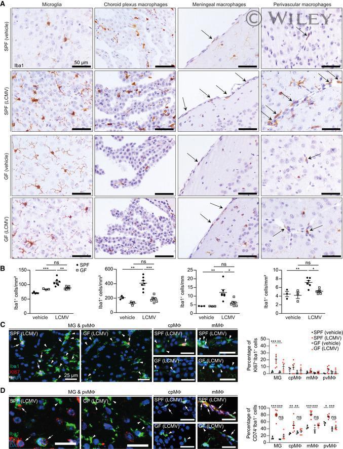
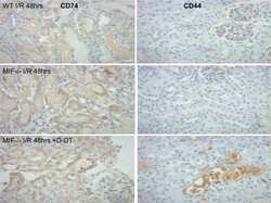
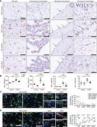
- 6 Figure Reduced immune response of myeloid cells to LCMV infection under GF conditions A Iba1 immunohistochemistry depicting cortical microglia, meningeal macrophages, perivascular macrophages, and choroid plexus macrophages 4 days after i.c. challenge with LCMV or vehicle controls. Arrows indicate Iba1 + meningeal or perivascular macrophages, respectively. B Quantification of Iba1 + microglia, choroid plexus macrophages, meningeal macrophages, and perivascular macrophages. Each symbol represents one mouse. Three to four sections per mouse were examined. Significant differences were evaluated by two-way ANOVA followed by Tukey's post hoc comparison test and marked with asterisks (* P < 0.05, ** P < 0.01, *** P < 0.001). Data are representative of two independent experiments. C, D Representative immunofluorescence images (left) of (C) Iba1 (green) and KI67 (red) or (D) CD74 (red) on cortical sections from SPF and GF mice. Nuclei were stained with DAPI (blue). Scale bars: 25 mum. White arrows indicate KI67 + /Iba1 + or CD74 + /Iba1 + macrophages, and arrowheads show KI67 - /Iba1 + or CD74 - /Iba1 + macrophages. Quantification thereof (right). Each symbol represents one mouse. At least three slides were examined per individual mouse. Data are presented as mean +- s.e.m. Significant differences were evaluated by two-way ANOVA followed by Tukey's post hoc comparison test and marked with asterisks (* P < 0.05, ** P < 0.01, *** P < 0.001). Data are representative of two independent