Explore
 Validate
 Learn
Learn Western blot
Antibody data
- Antibody Data
- Antigen structure
- References [3]
- Comments [0]
- Validations
- Western blot [1]
- Immunohistochemistry [1]
Submit
Validation data
Reference
Comment
Report error
- Product number
- MAB2417 - Provider product page

- Provider
- Novus Biologicals
- Product name
- Mouse Monoclonal Olig1 Antibody
- Antibody type
- Monoclonal
- Description
- Protein A or G purified from hybridoma culture supernatant. Detects human and mouse Olig1. In direct ELISAs, this antibody does not cross-react with recombinant human (rh) Olig2 or rhOlig3.
- Reactivity
- Human, Mouse
- Host
- Mouse
- Isotype
- IgG
- Vial size
- 100 ug
- Concentration
- LYOPH
- Storage
- Use a manual defrost freezer and avoid repeated freeze-thaw cycles. 12 months from date of receipt, -20 to -70 degreesC as supplied. 1 month, 2 to 8 degreesC under sterile conditions after reconstitution. 6 months, -20 to -70 degreesC under sterile conditions after reconstitution.
Submitted references Shortening the Half-Life of Cas9 Maintains Its Gene Editing Ability and Reduces Neuronal Toxicity.
Demyelination causes adult CNS progenitors to revert to an immature state and express immune cues that support their migration.
Reduced Morg1 expression in ischemic human brain.
Yang S, Li S, Li XJ
Cell reports 2018 Dec 4;25(10):2653-2659.e3
Cell reports 2018 Dec 4;25(10):2653-2659.e3
Demyelination causes adult CNS progenitors to revert to an immature state and express immune cues that support their migration.
Moyon S, Dubessy AL, Aigrot MS, Trotter M, Huang JK, Dauphinot L, Potier MC, Kerninon C, Melik Parsadaniantz S, Franklin RJ, Lubetzki C
The Journal of neuroscience : the official journal of the Society for Neuroscience 2015 Jan 7;35(1):4-20
The Journal of neuroscience : the official journal of the Society for Neuroscience 2015 Jan 7;35(1):4-20
Reduced Morg1 expression in ischemic human brain.
Haase D, Keiner S, Mawrin C, Wolf G
Neuroscience letters 2009 May 8;455(1):46-50
Neuroscience letters 2009 May 8;455(1):46-50
No comments: Submit comment
Supportive validation
- Submitted by
- Novus Biologicals (provider)
- Main image
- Experimental details
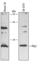
- Detection of Human Olig1 by Western Blot. Western blot shows lysates of Neuro-2A mouse neuroblastoma cell line and SH-SY5Y human neuroblastoma cell line. PVDF membrane was probed with 0.5 µg/mL of Human Olig1 Monoclonal Antibody (Catalog # MAB2417) followed by HRP-conjugated Anti-Mouse IgG Secondary Antibody (Catalog # HAF007). A specific band was detected for Olig1 at approximately 28 kDa (as indicated). This experiment was conducted under reducing conditions and using Immunoblot Buffer Group 1.

Supportive validation
- Submitted by
- Novus Biologicals (provider)
- Main image

- Experimental details
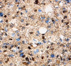
- Olig1 in Human Glioma. Olig1 was detected in immersion fixed paraffin-embedded sections of human glioma using 8 µg/mL Human Olig1 Monoclonal Antibody (Catalog # MAB2417) overnight at 4 °C. Tissue was stained with the Anti-Mouse HRP-DAB Cell & Tissue Staining Kit (brown; Catalog # CTS002) and counterstained with hematoxylin (blue). View our protocol for Chromogenic IHC Staining of Paraffin-embedded Tissue Sections.