Explore
 Validate
 Learn
Learn Flow cytometry
Antibody data
- Antibody Data
- Antigen structure
- References [2]
- Comments [0]
- Validations
- Flow cytometry [1]
- Other assay [2]
Submit
Validation data
Reference
Comment
Report error
- Product number
- 56-2239-42 - Provider product page

- Provider
- Invitrogen Antibodies
- Product name
- CD223 (LAG-3) Monoclonal Antibody (3DS223H), Alexa Fluor™ 700, eBioscience™
- Antibody type
- Monoclonal
- Antigen
- Other
- Description
- Description: This 3DS223H monoclonal antibody recognizes human CD223 also known as Lymphocyte Activation Gene 3 (LAG-3). LAG-3 is a 70-kDa surface glycoprotein belonging to the Ig superfamily with homology to CD4. LAG-3 binds to MHC class II with higher affinity than CD4 and is thought to be involved in the negative regulation of T cell activation and homeostatic proliferation. Surface expression of LAG-3 has been reported on activated T cells (including regulatory T cells) and NK cells. CD8+ T cells usually express LAG-3 at significantly higher levels than CD4+ T cells. Coexpression of LAG-3 and CD49b has been proposed to identify human and mouse Type 1 regulatory T cells (Tr1 cells).
- Conjugate
- Near infrared dye
- Antibody clone number
- 3DS223H
- Concentration
- 5 µL/Test
Submitted references SynNotch-CAR T cells overcome challenges of specificity, heterogeneity, and persistence in treating glioblastoma.
MAVS Genetic Variation Is Associated with Decreased HIV-1 Replication In Vitro and Reduced CD4(+) T Cell Infection in HIV-1-Infected Individuals.
Choe JH, Watchmaker PB, Simic MS, Gilbert RD, Li AW, Krasnow NA, Downey KM, Yu W, Carrera DA, Celli A, Cho J, Briones JD, Duecker JM, Goretsky YE, Dannenfelser R, Cardarelli L, Troyanskaya O, Sidhu SS, Roybal KT, Okada H, Lim WA
Science translational medicine 2021 Apr 28;13(591)
Science translational medicine 2021 Apr 28;13(591)
MAVS Genetic Variation Is Associated with Decreased HIV-1 Replication In Vitro and Reduced CD4(+) T Cell Infection in HIV-1-Infected Individuals.
Stunnenberg M, van Pul L, Sprokholt JK, van Dort KA, Gringhuis SI, Geijtenbeek TBH, Kootstra NA
Viruses 2020 Jul 16;12(7)
Viruses 2020 Jul 16;12(7)
No comments: Submit comment
Supportive validation
- Submitted by
- Invitrogen Antibodies (provider)
- Main image
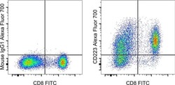
- Experimental details
- Normal human peripheral blood cells were stimulated for 3 days with Human IL-2 Recombinant Protein (Product # 14-8029-81), Anti-Human CD3, and Anti-Human CD28 Functional Grade Purifieds (Product # 16-0037-81 and Product # 16-0289-81). These cells were then surface stained with Anti-Human CD8a FITC (Product # 11-0087-42) and Mouse IgG1 K Isotype Control Alexa Fluor® 700 (Product # 56-4714-80) (left) or Anti-Human CD223 (LAG-3) Alexa Fluor® 700 (right). Viable cells in the lymphocyte gate, as determined by 7-AAD (Product # 00-6993-50), were used for analysis.
- Conjugate
- Near infrared dye

Supportive validation
- Submitted by
- Invitrogen Antibodies (provider)
- Main image
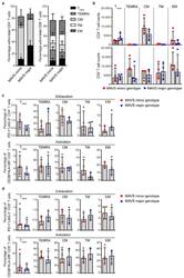
- Experimental details
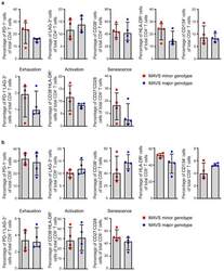
- Figure 3 MAVS genetic variation does not affect T cell exhaustion, activation, and senescence. ( a ) Percentages of PD-1 + , LAG-3 + , CD38 + , HLA-DR + , CD134 + , exhausted (PD-1 + LAG-3 + ), activated (CD38 + HLA-DR + ), and senescent (CD27 - CD28 - ) cells within CD4 + and ( b ) CD8 + T cells of untreated HIV-1-infected individuals with a MAVS minor or MAVS major genotype 2.5-3.5 years p.SC were analyzed using flow cytometry. Each square or dot represents a different study participant (median +- IQR). No significant differences between HIV-1-infected individuals with a MAVS minor or MAVS major genotype were observed.
- Conjugate
- Near infrared dye
- Submitted by
- Invitrogen Antibodies (provider)
- Main image
- Experimental details
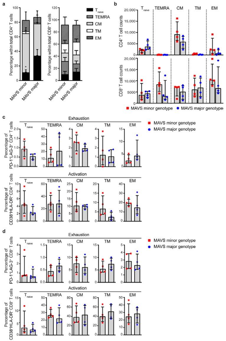
- Figure 4 MAVS minor genotype is associated with a decreased percentage of naive CD4 + T cells. ( a ) Percentages and ( b ) cell counts of naive (T naive ; CD45RA + CD27 + CCR7 + ), terminally differentiated effector memory (TEMRA; CD45RA + CCR7 - CD27 - ), central memory (CM; CD45RA - CCR7 + CD27 + ), transitional memory (TM; CD45RA - CCR7 - CD27 + ), and effector memory (EM; CD45RA - CCR7 - CD27 - ) cells within CD4 + and CD8 + T cells were analyzed using flow cytometry. ( c ) Percentages of exhausted (PD-1 + LAG-3 + ) and activated (CD38 + HLA-DR + ) CD4 + T cells and ( d ) CD8 + T cells within T naive , TEMRA, CM, TM, and EM populations were analyzed using flow cytometry. Each square or dot represents a different study participant (median +- IQR). All significant differences are indicated: * p < 0.05, unpaired Mann-Whitney test.
- Conjugate
- Near infrared dye