 Explore
 Validate
 Learn
Learn Flow cytometry
Antibody data
- Antibody Data
- Antigen structure
- References [8]
- Comments [0]
- Validations
- Flow cytometry [1]
- Other assay [2]
Submit
Validation data
Reference
Comment
Report error
- Product number
- 17-2769-42 - Provider product page

- Provider
- Invitrogen Antibodies
- Product name
- CD276 (B7-H3) Monoclonal Antibody (7-517), APC, eBioscience™
- Antibody type
- Monoclonal
- Antigen
- Other
- Description
- Description: This 7-517 monoclonal antibody reacts with human CD276, which is also known as B7-H3. This type I transmembrane protein is expressed on immature and mature dendritic cells but not monocytes, granulocytes, or resting lymphocytes. Moreover, CD276 is expressed on non-hematopoietic cells such as osteoblasts, fibroblasts, and epithelial cells. However, CD276 expression can be induced on dendritic cells, monocytes, T, B, and NK cells. This co-stimulatory molecule binds activated T cells, leading to cell proliferation and IFN-gamma production.
- Antibody clone number
- 7-517
- Concentration
- 5 µL/Test
Submitted references Methylome-based cell-of-origin modeling (Methyl-COOM) identifies aberrant expression of immune regulatory molecules in CLL.
Identification and validation of multiple cell surface markers of clinical-grade adipose-derived mesenchymal stromal cells as novel release criteria for good manufacturing practice-compliant production.
B7-H3 silencing by RNAi inhibits tumor progression and enhances chemosensitivity in U937 cells.
B7-H3 silencing inhibits tumor progression of mantle cell lymphoma and enhances chemosensitivity.
B7-H3 enhances tumor immunity in vivo by costimulating rapid clonal expansion of antigen-specific CD8+ cytolytic T cells.
Molecular characterization of human 4Ig-B7-H3, a member of the B7 family with four Ig-like domains.
The immune regulatory protein B7-H3 promotes osteoblast differentiation and bone mineralization.
B7-H3: a costimulatory molecule for T cell activation and IFN-gamma production.
Wierzbinska JA, Toth R, Ishaque N, Rippe K, Mallm JP, Klett LC, Mertens D, Zenz T, Hielscher T, Seifert M, Küppers R, Assenov Y, Lutsik P, Stilgenbauer S, Roessner PM, Seiffert M, Byrd J, Oakes CC, Plass C, Lipka DB
Genome medicine 2020 Mar 18;12(1):29
Genome medicine 2020 Mar 18;12(1):29
Identification and validation of multiple cell surface markers of clinical-grade adipose-derived mesenchymal stromal cells as novel release criteria for good manufacturing practice-compliant production.
Camilleri ET, Gustafson MP, Dudakovic A, Riester SM, Garces CG, Paradise CR, Takai H, Karperien M, Cool S, Sampen HJ, Larson AN, Qu W, Smith J, Dietz AB, van Wijnen AJ
Stem cell research & therapy 2016 Aug 11;7(1):107
Stem cell research & therapy 2016 Aug 11;7(1):107
B7-H3 silencing by RNAi inhibits tumor progression and enhances chemosensitivity in U937 cells.
Zhang W, Wang J, Wang Y, Dong F, Zhu M, Wan W, Li H, Wu F, Yan X, Ke X
OncoTargets and therapy 2015;8:1721-33
OncoTargets and therapy 2015;8:1721-33
B7-H3 silencing inhibits tumor progression of mantle cell lymphoma and enhances chemosensitivity.
Zhang W, Wang Y, Wang J, Dong F, Zhu M, Wan W, Li H, Wu F, Yan X, Ke X
International journal of oncology 2015;46(6):2562-72
International journal of oncology 2015;46(6):2562-72
B7-H3 enhances tumor immunity in vivo by costimulating rapid clonal expansion of antigen-specific CD8+ cytolytic T cells.
Luo L, Chapoval AI, Flies DB, Zhu G, Hirano F, Wang S, Lau JS, Dong H, Tamada K, Flies AS, Liu Y, Chen L
Journal of immunology (Baltimore, Md. : 1950) 2004 Nov 1;173(9):5445-50
Journal of immunology (Baltimore, Md. : 1950) 2004 Nov 1;173(9):5445-50
Molecular characterization of human 4Ig-B7-H3, a member of the B7 family with four Ig-like domains.
Steinberger P, Majdic O, Derdak SV, Pfistershammer K, Kirchberger S, Klauser C, Zlabinger G, Pickl WF, Stöckl J, Knapp W
Journal of immunology (Baltimore, Md. : 1950) 2004 Feb 15;172(4):2352-9
Journal of immunology (Baltimore, Md. : 1950) 2004 Feb 15;172(4):2352-9
The immune regulatory protein B7-H3 promotes osteoblast differentiation and bone mineralization.
Suh WK, Wang SX, Jheon AH, Moreno L, Yoshinaga SK, Ganss B, Sodek J, Grynpas MD, Mak TW
Proceedings of the National Academy of Sciences of the United States of America 2004 Aug 31;101(35):12969-73
Proceedings of the National Academy of Sciences of the United States of America 2004 Aug 31;101(35):12969-73
B7-H3: a costimulatory molecule for T cell activation and IFN-gamma production.
Chapoval AI, Ni J, Lau JS, Wilcox RA, Flies DB, Liu D, Dong H, Sica GL, Zhu G, Tamada K, Chen L
Nature immunology 2001 Mar;2(3):269-74
Nature immunology 2001 Mar;2(3):269-74
No comments: Submit comment
Supportive validation
- Submitted by
- Invitrogen Antibodies (provider)
- Main image
- Experimental details
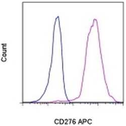
- Staining of normal human monocyte-derived dendritic cells with Mouse IgG1 K Isotype Control APC (Product # 17-4714-81) (blue histogram) or Anti-Human CD276 APC (purple histogram). Total viable cells were used for analysis.

Supportive validation
- Submitted by
- Invitrogen Antibodies (provider)
- Main image

- Experimental details
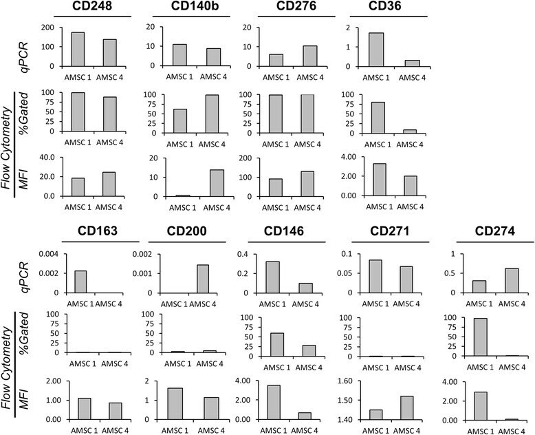
- Fig. 4 Expression of novel markers by quantitative PCR ( qPCR ) and flow cytometry. Gene expression data were compared to flow cytometry data for two donors [adipose-derived mesenchymal cell ( AMSC ) donors 1 and 4]. Highly abundant markers showed good concordance ( top panel ) between the techniques, whereas lower abundance markers showed variability ( bottom panel ). In particular, CD200 and CD274 were not correlated. MFI mean fluorescence intensity
- Submitted by
- Invitrogen Antibodies (provider)
- Main image
- Experimental details
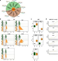
- Fig. 6 Flow cytometry analysis of T cell-/lymphocyte-specific markers on normal and malignant B cells from CLL patients. a Summary scheme representing functional implications of CLL-specific candidate genes selected for flow cytometric analysis. b Flow cytometric analysis of expression of CTLA-4, TIGIT, CD276, LILRB4, and CD2 on peripheral blood B cells of CLL patients. The expression was determined for non-malignant B cells (""Normal""; CD19 + CD5 - B cells, represented in green) and neoplastic B cells (""CLL"", CD19 + CD5 + B cells, represented in orange) detected in the same samples. ""Co,"" no antibody staining control; ""Ab,"" staining with the antibody of interest as indicated. c Normalized median fluorescence intensities (target MFI - MFI of negative control [Co]; nMFI). d Delta normalized median fluorescence intensities between CLL cells and normal B cells (DeltanMFI (CLL-normal)) for each patient tested
