Explore
 Validate
 Learn
Learn Flow cytometry
Antibody data
- Antibody Data
- Antigen structure
- References [1]
- Comments [0]
- Validations
- Flow cytometry [2]
Submit
Validation data
Reference
Comment
Report error
- Product number
- FAB2859N-025 - Provider product page

- Provider
- R&D Systems
- Product name
- Human Siglec-6/CD327 Alexa Fluor® 700-conjugated Antibody
- Antibody type
- Monoclonal
- Description
- Protein A or G purified from hybridoma culture supernatant. Detects human Siglec-6/CD327 in direct ELISAs.
- Reactivity
- Human
- Host
- Mouse
- Conjugate
- Near infrared dye
- Antigen sequence
NP_942142- Isotype
- IgG
- Antibody clone number
- 767329
- Vial size
- 25 Tests
- Concentration
- 0.2 mg/ml
- Storage
- Protect from light. Do not freeze. 12 months from date of receipt, 2 to 8 °C as supplied.
Submitted references Lipopolysaccharide inhalation recruits monocytes and dendritic cell subsets to the alveolar airspace.
Jardine L, Wiscombe S, Reynolds G, McDonald D, Fuller A, Green K, Filby A, Forrest I, Ruchaud-Sparagano MH, Scott J, Collin M, Haniffa M, Simpson AJ
Nature communications 2019 Apr 30;10(1):1999
Nature communications 2019 Apr 30;10(1):1999
No comments: Submit comment
Supportive validation
- Submitted by
- R&D Systems (provider)
- Main image

- Experimental details
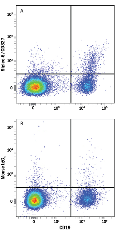
- Detection of Siglec-6/CD327 in Human PBMCs by Flow Cytometry. Human peripheral blood mononuclear cells (PBMCs) were stained with Mouse Anti-Human CD19 PE-conjugated Monoclonal Antibody (Catalog # FAB4867P) and either (A) Mouse Anti-Human Siglec-6/CD327 Alexa Fluor® 700-conjugated Monoclonal Antibody (Catalog # FAB2859N) or (B) Mouse Alexa Fluor 700 Isotype Control. View our protocol for Staining Membrane-associated Proteins.
- Submitted by
- R&D Systems (provider)
- Main image
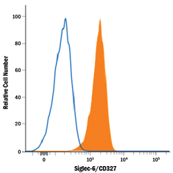
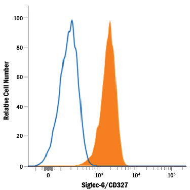
- Experimental details
- Detection of Siglec-6/CD327 in U937 Human Cell Line by Flow Cytometry. U937 human histiocytic lymphoma cell line was stained with Mouse Anti-Human Siglec-6/CD327 Alexa Fluor® 700-conjugated Monoclonal Antibody (Catalog # FAB2859N, filled histogram) or isotype control antibody (open histogram). View our protocol for Staining Membrane-associated Proteins.