Explore
 Validate
 Learn
Learn720387
antibody from Invitrogen Antibodies
Targeting: HMGN1
FLJ27265, FLJ31471, HMG14, MGC104230, MGC117425
 Western blot
Antibody data
- Antibody Data
- Antigen structure
- References [0]
- Comments [0]
- Validations
- Western blot [3]
- Immunocytochemistry [1]
- Chromatin Immunoprecipitation [1]
Submit
Validation data
Reference
Comment
Report error
- Product number
- 720387 - Provider product page

- Provider
- Invitrogen Antibodies
- Product name
- HMGN1 Polyclonal Antibody
- Antibody type
- Polyclonal
- Antigen
- Synthetic peptide
- Description
- This antibody predicted to react with Monkey, Bovine, Sheep
- Reactivity
- Human
- Host
- Rabbit
- Isotype
- IgG
- Vial size
- 100 µg
- Concentration
- 0.5 mg/mL
- Storage
- Store at 4°C short term. For long term storage, store at -20°C, avoiding freeze/thaw cycles.
No comments: Submit comment
Supportive validation
- Submitted by
- Invitrogen Antibodies (provider)
- Main image

- Experimental details
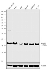
- Western blot analysis was performed on Modified Whole cell extracts (1% SDS) (30 µg lysate) of MDA-MB-231 (Lane 1), T-47D (Lane 2), A549 (Lane 3), Hep G2 (Lane 4), NTERA-2 (Lane 5) and NCCIT (Lane 6). The blots were probed with Anti-HMGN1 Rabbit Polyclonal Antibody (Product # 720387, 2 µg/mL) and detected by chemiluminescence using Goat anti-Rabbit IgG (H+L) Superclonal™ Secondary Antibody, HRP conjugate (Product # A27036, 0.2 µg/mL, 1:5000 dilution). A 17 kDa band corresponding to HMGN1 was observed across the cell lines tested. Known quantity of protein samples were electrophoresed using Novex® NuPAGE® 10% Bis-Tris gel (Product # NP0302BOX), XCell SureLock™ Electrophoresis System (Product # EI0002) and Novex® Sharp Pre-Stained Protein Standard (Product # LC5800). Resolved proteins were then transferred onto a nitrocellulose membrane with iBlot® Dry Blotting System (Product # IB21001). The membrane was probed with the relevant primary and secondary Antibody following blocking with 5% skimmed milk. Chemiluminescent detection was performed using Pierce™ ECL Western Blotting Substrate (Product # 32106).

- Submitted by
- Invitrogen Antibodies (provider)
- Main image

- Experimental details
- Western blot analysis was performed on Modified Whole cell extracts (1% SDS) (30 µg lysate) of MDA-MB-231 (Lane 1), T-47D (Lane 2), A549 (Lane 3), Hep G2 (Lane 4), NTERA-2 (Lane 5) and NCCIT (Lane 6). The blots were probed with Anti-HMGN1 Rabbit Polyclonal Antibody (Product # 720387, 2 µg/mL) and detected by chemiluminescence using Goat anti-Rabbit IgG (H+L) Superclonal™ Secondary Antibody, HRP conjugate (Product # A27036, 0.2 µg/mL, 1:5000 dilution). A 17 kDa band corresponding to HMGN1 was observed across the cell lines tested. Known quantity of protein samples were electrophoresed using Novex® NuPAGE® 10% Bis-Tris gel (Product # NP0302BOX), XCell SureLock™ Electrophoresis System (Product # EI0002) and Novex® Sharp Pre-Stained Protein Standard (Product # LC5800). Resolved proteins were then transferred onto a nitrocellulose membrane with iBlot® Dry Blotting System (Product # IB21001). The membrane was probed with the relevant primary and secondary Antibody following blocking with 5% skimmed milk. Chemiluminescent detection was performed using Pierce™ ECL Western Blotting Substrate (Product # 32106).

- Submitted by
- Invitrogen Antibodies (provider)
- Main image
- Experimental details
- Western blot analysis was performed on Modified Whole cell extracts (1% SDS) (30 µg lysate) of MDA-MB-231 (Lane 1), T-47D (Lane 2), A549 (Lane 3), Hep G2 (Lane 4), NTERA-2 (Lane 5) and NCCIT (Lane 6). The blots were probed with Anti-HMGN1 Rabbit Polyclonal Antibody (Product # 720387, 2 µg/mL) and detected by chemiluminescence using Goat anti-Rabbit IgG (H+L) Superclonal™ Secondary Antibody, HRP conjugate (Product # A27036, 0.2 µg/mL, 1:5000 dilution). A 17 kDa band corresponding to HMGN1 was observed across the cell lines tested. Known quantity of protein samples were electrophoresed using Novex® NuPAGE® 10% Bis-Tris gel (Product # NP0302BOX), XCell SureLock™ Electrophoresis System (Product # EI0002) and Novex® Sharp Pre-Stained Protein Standard (Product # LC5800). Resolved proteins were then transferred onto a nitrocellulose membrane with iBlot® Dry Blotting System (Product # IB21001). The membrane was probed with the relevant primary and secondary Antibody following blocking with 5% skimmed milk. Chemiluminescent detection was performed using Pierce™ ECL Western Blotting Substrate (Product # 32106).

Supportive validation
- Submitted by
- Invitrogen Antibodies (provider)
- Main image
- Experimental details
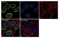
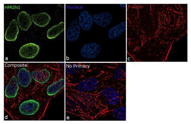
- For immunofluorescence analysis, HeLa cells were fixed and permeabilized for detection of endogenous HMGN1 using Anti- HMGN1 Rabbit Polyclonal Antibody (Product # 720387, 5 µg/mL) and labeled with Goat anti-Rabbit IgG (H+L) Superclonal™ Secondary Antibody, Alexa Fluor® 488 conjugate (Product # A27034, 1:2000). Panel a) shows representative cells that were stained for detection and localization of HMGN1 protein (green), Panel b) is stained for nuclei (blue) using SlowFade® Gold Antifade Mountant with DAPI (Product # S36938). Panel c) represents cytoskeletal F-actin staining using Rhodamine Phalloidin (Product # R415, 1:300). Panel d) is a composite image of Panels a, b and c clearly demonstrating Nuclear localization of HMGN1. Panel e) represents control cells with no primary antibody to assess background. The images were captured at 60X magnification.
Supportive validation
- Submitted by
- Invitrogen Antibodies (provider)
- Main image
- Experimental details
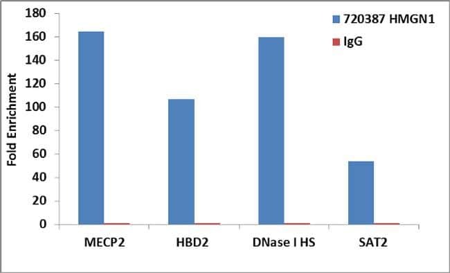
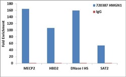
- Enrichment of endogenous HMGN1 protein at specific gene loci using Anti-HMGN1 Rabbit Polyclonal Antibody: Chromatin Immunoprecipitation (ChIP) was performed using Anti-HMGN1 Rabbit Polyclonal Antibody (Product # 720387, 3 µg) on sheared chromatin from 2 million HeLa cells using the MAGnify ChIP system kit (Product # 49-2024). Normal Rabbit IgG (1 µg) was used as a negative IP control. The purified DNA was analyzed by 7500 Fast qPCR system (Product # 4351106) with optimized PCR primer pairs for the promoters of the active MECP2, HBD2, DNase I Hypersensitive region used as positive control target genes, and the region of the inactive SAT2 satellite repeat, used as negative control target gene. Data is presented as fold enrichment of the antibody signal versus the negative control IgG using the comparative CT method.