Explore
 Validate
 Learn
Learn Western blot
 Flow cytometry
Antibody data
- Antibody Data
- Antigen structure
- References [0]
- Comments [0]
- Validations
- Western blot [5]
- Immunocytochemistry [3]
- Immunohistochemistry [3]
Submit
Validation data
Reference
Comment
Report error
- Product number
- MA5-25120 - Provider product page

- Provider
- Invitrogen Antibodies
- Product name
- Profilin 1 Monoclonal Antibody (OTI2D2)
- Antibody type
- Monoclonal
- Antigen
- Recombinant full-length protein
- Reactivity
- Human, Rat, Canine
- Host
- Mouse
- Isotype
- IgG
- Antibody clone number
- OTI2D2
- Vial size
- 100 µL
- Concentration
- 0.58 mg/mL
- Storage
- -20° C, Avoid Freeze/Thaw Cycles
No comments: Submit comment
Supportive validation
- Submitted by
- Invitrogen Antibodies (provider)
- Main image
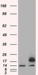
- Experimental details
- Western blot analysis of PFN1 in HEK293T cells in untransfected (Left lane) and transfected (Right lane) samples using 5 µg per lane. The samples were separated by SDS-PAGE and probed with PFN1 (Product # MA5-25120) monoclonal antibody.
- Submitted by
- Invitrogen Antibodies (provider)
- Main image

- Experimental details
- Western blot analysis of PFN1 in HepG2, HeLa, HT29, A549, COS7, Jurkat, MDCK, PC12, MCF7 cells using 35 µg per lane. Samples were probed with PFN1 (Product # MA5-25120) monoclonal antibody.
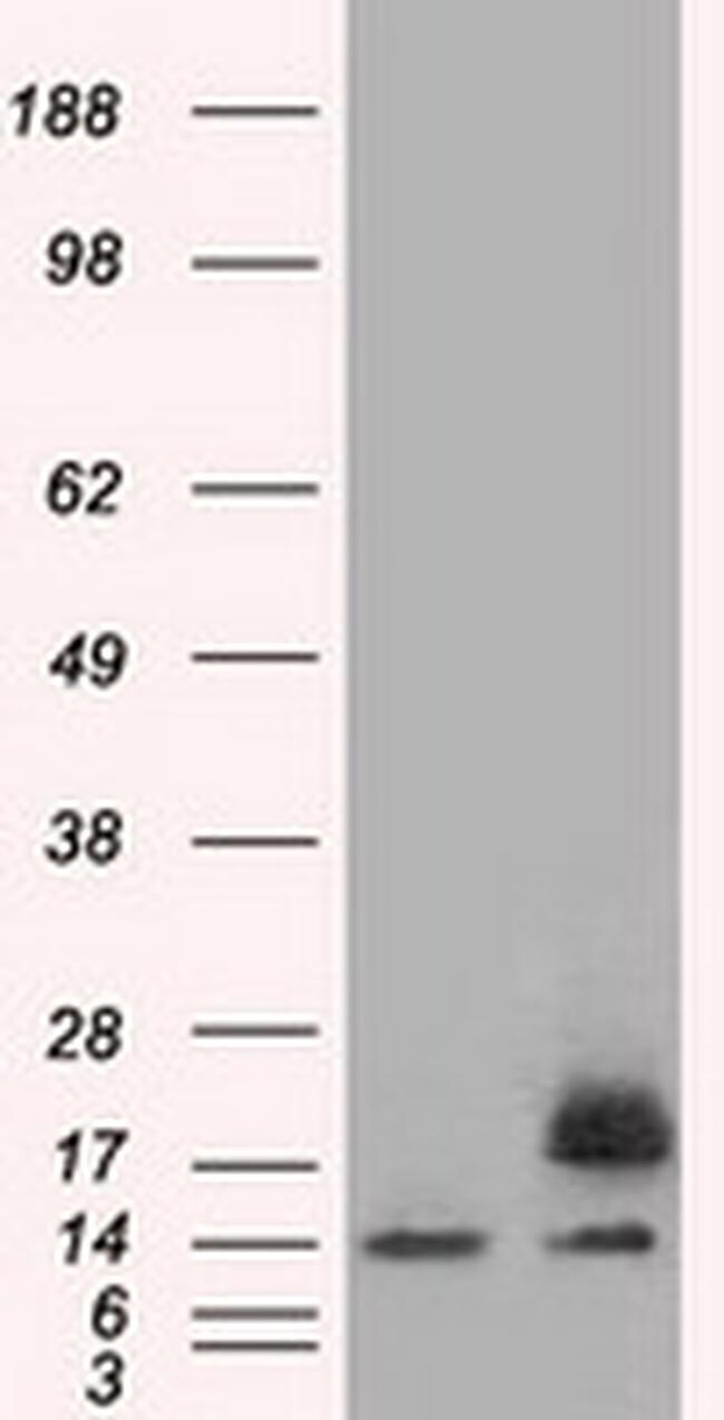
- Submitted by
- Invitrogen Antibodies (provider)
- Main image
- Experimental details
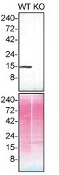
- Western blot of Profilin 1 (PFN1) was performed by loading 50 µg of HAP1 WT (lane 1) and PFN1 KO (lane 2) cell lysates onto a NuPAGE™ 10%, Bis-Tris, 1.0 mm, Midi Protein Gel (Product # WG1201BOX). Proteins on the blot were visualized with Ponceau staining (below immunoblot). Proteins were transferred to nitrocellulose membrane and blocked in 5% milk for 1 hr. PFN1 was detected at approximately 15 kDa using Profilin 1 Monoclonal Antibody (OTI2D2) (Product # MA5-25120) at a dilution of 1:2000 in 5% BSA in TBST overnight at 4 degree celsius. The blot was probed with Goat anti-Mouse IgG (H+L) Secondary Antibody, HRP (Product # 62-6520) diluted to 0.2 µg/mL in TBST with 5% milk for 1 hr at room temperature. Chemiluminescent detection was performed using ECL Western Blotting Substrate. Data courtesy of YCharOS Inc., an open science company with the mission of characterizing commercially available antibodies using knockout validation.

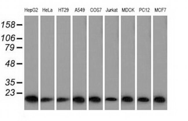
- Submitted by
- Invitrogen Antibodies (provider)
- Main image

- Experimental details
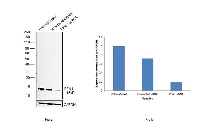
- Knockdown of Profilin 1 was achieved by transfecting HeLa cells with Profilin 1 specific siRNAs (Silencer® select Product # s10375, s10376 ). Western blot analysis (Fig. a) was performed using Whole cell extracts from the Profilin 1 knockdown cells (lane 3), non-targeting scrambled siRNA transfected cells (lane 2) and untransfected cells (lane 1). The blot was probed with Profilin 1 Monoclonal Antibody (OTI2D2) (Product # MA5-25120, 1:1000 dilution) and Goat anti-Mouse IgG (H+L) Superclonal™ Recombinant Secondary Antibody, HRP (Product # A28177, 1:4000 dilution). Densitometric analysis of this western blot is shown in histogram (Fig. b). Decrease in signal upon siRNA mediated knock down confirms that antibody is specific to Profilin 1.
- Submitted by
- Invitrogen Antibodies (provider)
- Main image
- Experimental details
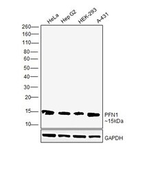
- Western blot was performed using Anti-Profilin 1 Monoclonal Antibody (OTI2D2)(Product # MA5-25120) and a 15kDa band corresponding to Profilin 1 was observed across cell lines tested. Whole cell extracts (30 µg lysate) of HeLa (Lane 1), Hep G2 (Lane 2), HEK-293 (Lane 3) and A-431 (Lane 4) were electrophoresed using NuPAGE™ 12% Bis-Tris Protein Gel (Product # NP0341BOX). Resolved proteins were then transferred onto a Nitrocellulose membrane (Product # IB23001) by iBlot® 2 Dry Blotting System (Product # IB21001). The blot was probed with the primary antibody (1:1500 dilution) and detected by chemiluminescence with Goat anti-Mouse IgG (H+L) Superclonal™ Recombinant Secondary Antibody, HRP (Product # A28177, 1:4000 dilution) using the iBright FL 1000 (Product # A32752). Chemiluminescent detection was performed using Novex® ECL Chemiluminescent Substrate Reagent Kit (Product # WP20005).

Supportive validation
- Submitted by
- Invitrogen Antibodies (provider)
- Main image

- Experimental details
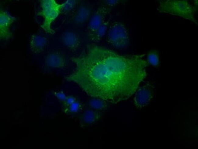
- Immunofluorescent analysis of PFN1 in COS7 cells. Cells were transfected with a plasmid overexpressing PFN1 and probed with a PFN1 monoclonal antibody (Product # MA5-25120).
- Submitted by
- Invitrogen Antibodies (provider)
- Main image

- Experimental details
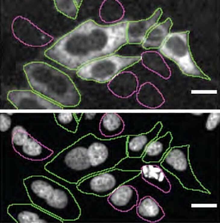
- Immunofluorescence of Profilin 1 (PFN1) was performed using HAP1 WT and PFN1 KO cells that were transfected with a green or a far-red fluorescent dye, respectively. Post-transfection, WT and KO cells were mixed and plated to a 1:1 ratio in a 96-well plate with glass bottom as a mosaic and incubated for 24 hrs. Cells were fixed in 4% PFA (in PBS) for 15 min at RT; cells were permeabilized with 0.1% Triton X-100 for 10 min at RT and blocked with PBS with 5% BSA, 5% goat serum, and 0.01% Triton X-100 for 30 min. Cells were stained with Profilin 1 Monoclonal Antibody (OTI2D2) (Product # MA5-25120) at a 1.0 μg/mL overnight at 4 degree celcius. Secondary antibody incubation was performed using 1 µg/mL of Goat anti-Mouse IgG (H+L) Highly Cross-Adsorbed Secondary Antibody, Alexa Fluor™ 555 (Product # A-21424) together with DAPI for 1 hr at RT. Imaging was performed with a 20X water immersion objective. Representative images where WT and KO cells are outlined with a green (WT) or magenta (KO) line, respectively, are shown. The top and bottom panels show antibody and DAPI stainings, respectively. Scale bar = 10 μm. Data courtesy of YCharOS Inc., an open science company with the mission of characterizing commercially available antibodies using knockout validation.
- Submitted by
- Invitrogen Antibodies (provider)
- Main image
- Experimental details
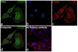
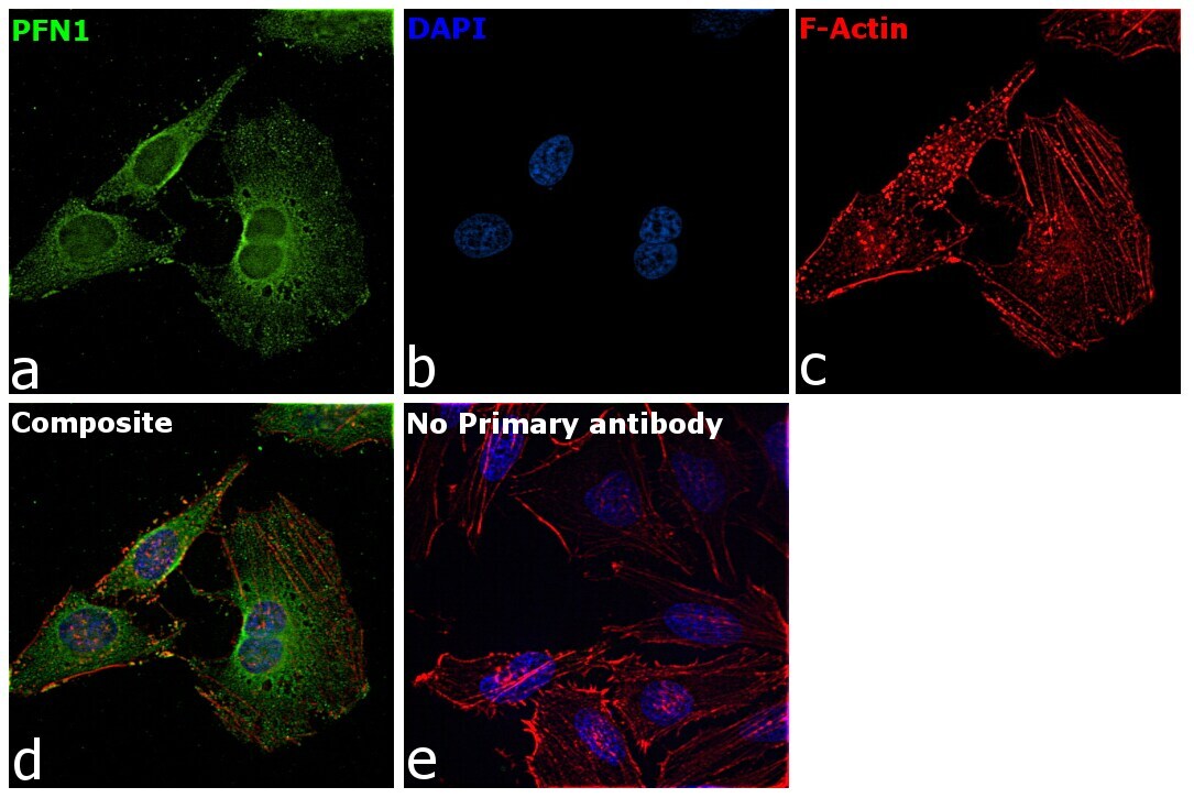
- Immunofluorescence analysis of Profilin 1 was performed using 70% confluent log phase HeLa cells. The cells were fixed with 4% paraformaldehyde for 10 minutes, permeabilized with 0.1% Triton™ X-100 for 15 minutes, and blocked with 2% BSA for 45 minutes at room temperature. The cells were labeled with Profilin 1 Monoclonal Antibody (OTI2D2) (Product # MA5-25120) at 1:100 dilution in 0.1% BSA, incubated at 4 degree celsius overnight and then labeled with Goat anti-Mouse IgG (H+L) Superclonal™ Recombinant Secondary Antibody, Alexa Fluor® 488 conjugate (Product # A28175), (1:2000 dilution), for 45 minutes at room temperature (Panel a: Green). Nuclei (Panel b:Blue) were stained with ProLong™ Diamond Antifade Mountant with DAPI (Product # P36962). F-actin (Panel c: Red) was stained with Rhodamine Phalloidin (Product # R415, 1:300 dilution). Panel d represents the merged image showing cytoskeleton localization. Panel e represents control cells with no primary antibody to assess background. The images were captured at 60X magnification.
Supportive validation
- Submitted by
- Invitrogen Antibodies (provider)
- Main image
- Experimental details
- Immunohistochemistry was performed on paraffin-embedded human kidney tissue. To expose target proteins, 10mM citric buffer, pH6.0, 100°C for 10min was used. Following antigen retrieval, tissues were probed with a PFN1 monoclonal antibody (Product # MA5-25120) at a dilution of 1:50.

- Submitted by
- Invitrogen Antibodies (provider)
- Main image
- Experimental details
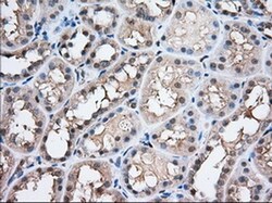
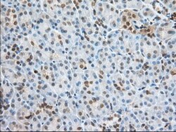
- Immunohistochemistry was performed on paraffin-embedded human pancreas tissue. To expose target proteins, 10mM citric buffer, pH6.0, 100°C for 10min was used. Following antigen retrieval, tissues were probed with a PFN1 monoclonal antibody (Product # MA5-25120) at a dilution of 1:50.

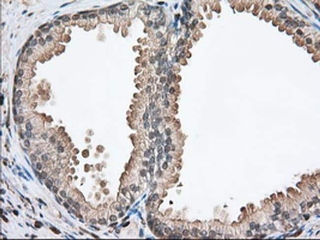
- Submitted by
- Invitrogen Antibodies (provider)
- Main image

- Experimental details
- Immunohistochemistry was performed on paraffin-embedded human prostate tissue. To expose target proteins, 10mM citric buffer, pH6.0, 100°C for 10min was used. Following antigen retrieval, tissues were probed with a PFN1 monoclonal antibody (Product # MA5-25120) at a dilution of 1:50.