Explore
 Validate
 Learn
Learn Western blot
Antibody data
- Antibody Data
- Antigen structure
- References [1]
- Comments [0]
- Validations
- Western blot [1]
- Immunocytochemistry [1]
- Chromatin Immunoprecipitation [1]
- Other assay [1]
Submit
Validation data
Reference
Comment
Report error
- Product number
- 720166 - Provider product page

- Provider
- Invitrogen Antibodies
- Product name
- Histone H4 Polyclonal Antibody
- Antibody type
- Polyclonal
- Antigen
- Other
- Description
- These Polyclonal antibodies are of rabbit origin developed by immunizing animals with proteins or peptides. The polyclonal antibody is purified by affinity purification from the rabbit sera generated after immunizing the rabbits with a specific type of protein or peptide. The purified antibody is tested for its functionality in various relevant research applications. The antibody is developed for Research Use Only and is non-hazardous or non-infectious in nature.
- Concentration
- 0.5 mg/mL
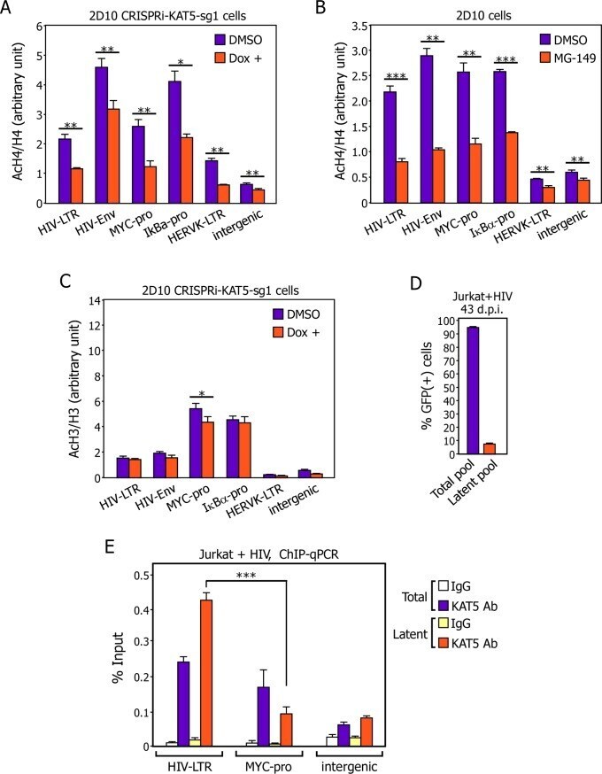
Submitted references The KAT5-Acetyl-Histone4-Brd4 axis silences HIV-1 transcription and promotes viral latency.
Li Z, Mbonye U, Feng Z, Wang X, Gao X, Karn J, Zhou Q
PLoS pathogens 2018 Apr;14(4):e1007012
PLoS pathogens 2018 Apr;14(4):e1007012
No comments: Submit comment
Supportive validation
- Submitted by
- Invitrogen Antibodies (provider)
- Main image
- Experimental details
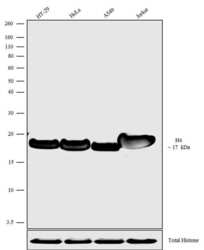
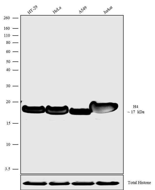
- Western blot analysis was performed on acid extracts (30 µg lysate) of HT-29 (Lane 1), HeLa (Lane 2), A549 (Lane 3) and Jurkat (Lane 4). The blots were probed with Anti-Histone H4 Rabbit Polyclonal Antibody (Product # 720166, 1-2 µg/mL) and detected by chemiluminescence using Goat anti-Rabbit IgG (H+L) Superclonal™ Secondary Antibody, HRP conjugate (Product # A27036, 0.4 µg/mL, 1:2500 dilution). A 17 kDa band corresponding to Histone H4 was observed. Known quantity of protein samples were electrophoresed using Novex® NuPAGE® 12% Bis-Tris gel (Product # NP0342BOX), XCell SureLock™ Electrophoresis System (Product # EI0002) and Novex® Sharp Pre-Stained Protein Standard (Product # LC5800). Resolved proteins were then transferred onto a nitrocellulose membrane with iBlot® Dry Blotting System (Product # IB21001). The membrane was probed with the relevant primary and secondary Antibody following blocking with 5% skimmed milk. Chemiluminescent detection was performed using Pierce™ ECL Western Blotting Substrate (Product # 32106).
Supportive validation
- Submitted by
- Invitrogen Antibodies (provider)
- Main image
- Experimental details
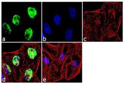
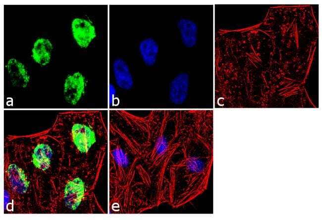
- Immunofluorescence was performed on fixed and permeabilized HeLa cells for detection of Histone H4 using Histone H4 Rabbit Polyclonal antibody (Product # 720166, 2 µg/mL) and labeled with Goat anti-Rabbit IgG (H+L) Superclonal™ Secondary Antibody, Alexa Fluor® 488 conjugate (Product # A27034, 1:2000). Panel a) shows representative cells that were stained for detection and localization of Histone H4 protein (green), Panel b) is stained for nuclei (blue) using SlowFade® Gold Antifade Mountant with DAPI (Product # S36938). Panel c) represents cytoskeletal F-actin staining using Alexa Fluor® 555 Rhodamine Phalloidin (Product # R415, 1:300). Panel d) is a composite image of Panels a, b and c clearly demonstrating nuclear localization of Histone H4. Panel e) represents control cells with no primary antibody to assess background.
Supportive validation
- Submitted by
- Invitrogen Antibodies (provider)
- Main image
- Experimental details
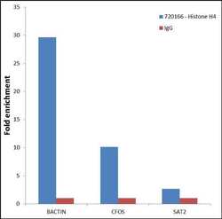
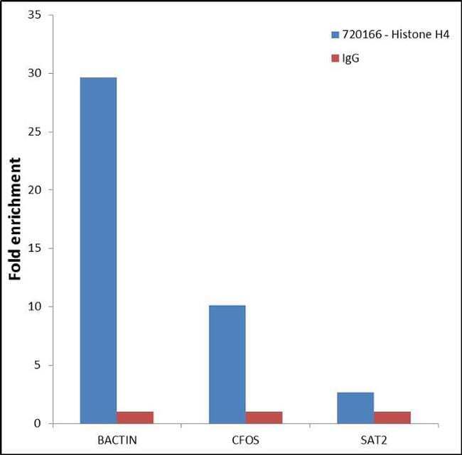
- Enrichment of endogenous Histone H4 protein using Anti- Histone H4 Rabbit Polyclonal Antibody: Chromatin Immunoprecipitation (ChIP) was performed using Anti- Histone H4 Rabbit Polyclonal Antibody (Product # 720166, 3 µg) on sheared chromatin from 2 million Jurkat cells using the "MAGnify ChIP system" kit (Product # 49-2024). Normal Rabbit IgG (1 µg) was used as a negative IP control. The purified DNA was analyzed by 7500 Fast qPCR system (Product # 4351106) with optimized PCR primer pairs for the promoters of the active cFOS, B-Actin genes, region used as positive control target gene, and the region of the inactive SAT2 satellite repeat, used as negative control target gene. Data is presented as fold enrichment of the antibody signal versus the negative control IgG using the comparative CT method.
Supportive validation
- Submitted by
- Invitrogen Antibodies (provider)
- Main image
- Experimental details
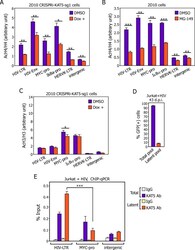
- Fig 4 Antagonizing KAT5 reduces AcH4 but not AcH3 level on both HIV and non-HIV gene promoters and a higher level of KAT5 exists on the viral LTR than on the cellular MYC gene promoter in latently infected cells. A., B., & C. The 2D10-based CRISPRi-KAT5-sg1 (A & C) and parental 2D10 (B) cells were treated with the indicated drugs and subjected to ChIP-qPCR assays to determine the levels of AcH3, AcH4, total H3 and total H4 bound to the various genomic locations labeled at the bottom. The ChIP-qPCR signals were normalized to those of input DNA for each genomic location and the ratios of AcH3 over H3 and AcH4 over H4 were shown. The error bars represent mean +/- SD from three independent qPCR reactions. The asterisks (*: p