 Explore
 Validate
 Learn
Learn Western blot
 Immunocytochemistry
Antibody data
- Antibody Data
- Antigen structure
- References [0]
- Comments [0]
- Validations
- Western blot [1]
- ELISA [1]
- Chromatin Immunoprecipitation [1]
Submit
Validation data
Reference
Comment
Report error
- Product number
- NBP2-59264 - Provider product page

- Provider
- Novus Biologicals
- Product name
- Rabbit Polyclonal Histone H4 Antibody
- Antibody type
- Polyclonal
- Description
- Peptide affinity purified.
- Reactivity
- Human
- Host
- Rabbit
- Isotype
- IgG
- Vial size
- 50 ug
- Storage
- Store at -20C. Avoid freeze-thaw cycles.
No comments: Submit comment
Supportive validation
- Submitted by
- Novus Biologicals (provider)
- Main image
- Experimental details
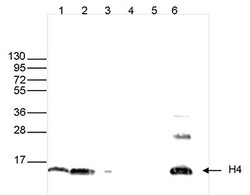
- Western Blot: Histone H4 Antibody [NBP2-59264] - Western blot was performed on whole cell extracts (25 ug, lane 1) and histone extracts (15 ug, lane 2) from HeLa cells, and on 1 ug of recombinant histone H2A, H2B, H3 and H4 (lane 3, 4, 5 and 6, respectively) using the antibody against H4 . The antibody was diluted 1:1,000 in TBS-Tween containing 5% skimmed milk. The position of the protein of interest is indicated on the right; the marker (in kDa) is shown on the left.

Supportive validation
- Submitted by
- Novus Biologicals (provider)
- Main image

- Experimental details
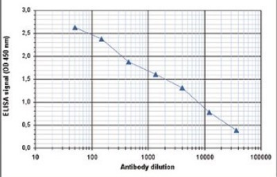
- ELISA: Histone H4 Antibody [NBP2-59264] - To determine the titer of the antibody, an ELISA was performed using a serial dilution of the antibody directed against H4 in antigen coated wells. By plotting the absorbance against the antibody dilution, the titer of the antibody was estimated to be 1:3,000.
Supportive validation
- Submitted by
- Novus Biologicals (provider)
- Main image
- Experimental details
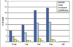
- Chromatin Immunoprecipitation: Histone H4 Antibody [NBP2-59264] - ChIP assays were performed using human HeLa cells, the antibody against H4 and optimized PCR primer pairs for qPCR. ChIP was performed using sheared chromatin from 1 million cells. A titration consisting of 0.5, 1, 2 and 5 ug of antibody per ChIP experiment was analyzed. IgG (1 ug/IP) was used as a negative IP control. Quantitative PCR was performed with primers specific for the promoters of the active GAPDH and c-fos genes, and for the inactive MYOD1 gene and the Sat2 satellite repeat. Figure shows the recovery, expressed as a % of input (the relative amount of immunoprecipitated DNA compared to input DNA after qPCR analysis).
