 Explore
 Validate
 Learn
LearnMAB1147-100
antibody from R&D Systems
Targeting: MYDGF
C19orf10, IL-25, IL-27, IL25, IL27, IL27w, R33729_1, SF20
 Western blot
Antibody data
- Antibody Data
- Antigen structure
- References [0]
- Comments [0]
- Validations
- Western blot [1]
- Flow cytometry [1]
Submit
Validation data
Reference
Comment
Report error
- Product number
- MAB1147-100 - Provider product page

- Provider
- R&D Systems
- Product name
- Human SF20/MYDGF Antibody
- Antibody type
- Monoclonal
- Description
- Protein A or G purified from hybridoma culture supernatant. Detects human SF20/MYDGF in direct ELISAs.
- Reactivity
- Human
- Host
- Mouse
- Conjugate
- Unconjugated
- Antigen sequence
Q969H8- Isotype
- IgG
- Antibody clone number
- 1009420
- Vial size
- 100 ug
- Storage
- Use a manual defrost freezer and avoid repeated freeze-thaw cycles. 12 months from date of receipt, -20 to -70 °C as supplied. 1 month, 2 to 8 °C under sterile conditions after reconstitution. 6 months, -20 to -70 °C under sterile conditions after reconstitution.
No comments: Submit comment
Supportive validation
- Submitted by
- R&D Systems (provider)
- Main image

- Experimental details
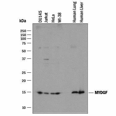
- Detection of Human SF20 by Western Blot. Western blot shows lysates of DU145 human prostate carcinoma cell line, Jurkat human acute T cell leukemia cell line, HeLa human cervical epithelial carcinoma cell line, WI-38 human lung fibroblast cell line, human lung tissue, and human liver tissue. PVDF membrane was probed with 2 µg/mL of Mouse Anti-Human SF20 Monoclonal Antibody (Catalog # MAB1147) followed by HRP-conjugated Anti-Mouse IgG Secondary Antibody (Catalog # HAF018). A specific band was detected for SF20 at approximately 18 kDa (as indicated). This experiment was conducted under reducing conditions and using Immunoblot Buffer Group 1.
Supportive validation
- Submitted by
- R&D Systems (provider)
- Main image

- Experimental details
- Detection of SF20 on U937 Human Cell Line by Flow Cytometry. U937 Human histiocytic lymphoma cell line was stained with Mouse Anti-Human SF20 Monoclonal Antibody (Catalog # MAB1147, filled histogram) or Mouse IgG1 isotype control antibody (Catalog # MAB002, open histogram) followed by APC-conjugated Anti-Mouse IgG Secondary Antibody (Catalog # F0101B). To facilitate intracellular staining, cells were fixed with paraformaldehyde and permeabilized with saponin. View our protocol for Staining Intracellular Molecules.
