Explore
 Validate
 Learn
Learn Western blot
Antibody data
- Antibody Data
- Antigen structure
- References [0]
- Comments [0]
- Validations
- Western blot [1]
- Immunohistochemistry [2]
Submit
Validation data
Reference
Comment
Report error
- Product number
- 711422 - Provider product page

- Provider
- Invitrogen Antibodies
- Product name
- CORIN Recombinant Polyclonal Antibody
- Antibody type
- Polyclonal
- Antigen
- Other
- Description
- This antibody is predicted to react with Monkey, Cat, Horse
- Concentration
- 0.5 mg/mL
No comments: Submit comment
Supportive validation
- Submitted by
- Invitrogen Antibodies (provider)
- Main image

- Experimental details
- Western blot analysis was performed on Tissue extracts (30 µg lysate) of Mouse Heart (Lane 1), Rat Heart (Lane 2), Mouse Brain (Lane 3), Rat Brain (Lane 4), Mouse Kidney (Lane 5) and Rat Kidney (Lane 6). The blots were probed with Anti-Corin Recombinant Rabbit Polyclonal Antibody (Product # 711422, 1-2 µg/mL) and detected by chemiluminescence using Goat anti-Rabbit IgG (H+L) Superclonal™ Secondary Antibody, HRP conjugate (Product # A27036, 0.4 µg/mL, 1:5000 dilution). A 220 kDa band corresponding to Corin was observed only in heart tissue extracts tested. Known quantity of protein samples were electrophoresed using Novex® NuPAGE® 4-12% Bis-Tris gel (Product # NP0321BOX), XCell SureLock™ Electrophoresis System (Product # EI0002) and Novex® Sharp Pre-Stained Protein Standard (Product # LC5800). Resolved proteins were then transferred onto a nitrocellulose membrane with wet transfer. The membrane was probed with the relevant primary and secondary Antibody following blocking with 5% skimmed milk. Chemiluminescent detection was performed using Pierce™ ECL Western blotting Substrate (Product # 32106).

Supportive validation
- Submitted by
- Invitrogen Antibodies (provider)
- Main image
- Experimental details
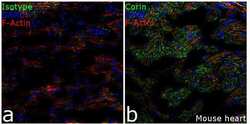
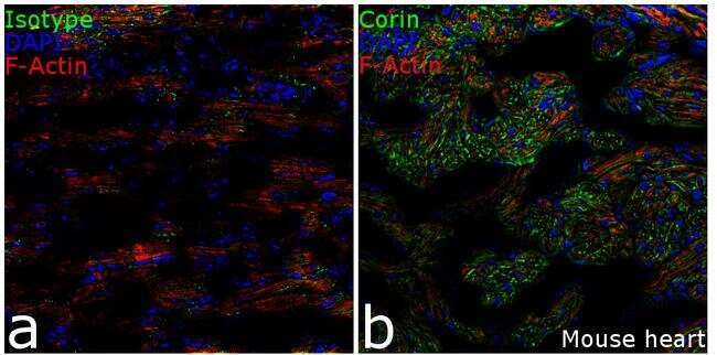
- Immunofluorescence analysis of Corin in mouse heart tissue: Frozen sections were fixed with 4% PFA for 20 min, permeabilized using 0.1% Triton-X 100 for 10 mins and blocked for 1 hour with 2% BSA. Transverse sections of mouse heart atrium were incubated with Anti- Corin Recombinant Rabbit Polyclonal Antibody (Product # 711422, 2 µg/mL) overnight at 4°C, followed by Goat anti-Rabbit IgG (H+L) Superclonal™ Secondary Antibody, Alexa Fluor® 488 conjugate (Product # A27034, 1:2000, 45 mins). Nuclei (blue) were stained using SlowFade® Gold Antifade Mountant with DAPI (Product # S36938), and cytoskeletal F-actin (red) was stained using Rhodamine Phalloidin (Product # R415, 1:300). Panel a) represents staining with the matched isotype control. Panel b) shows a representative section stained for Corin (green). The images were captured at 20X magnification.
- Submitted by
- Invitrogen Antibodies (provider)
- Main image
- Experimental details
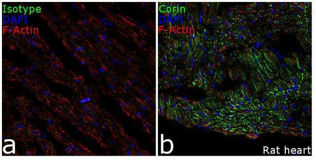
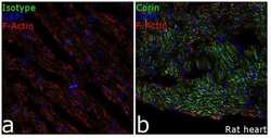
- Immunofluorescence analysis of Corin in rat heart tissue: Frozen sections were fixed with 4% PFA for 20 min, permeabilized using 0.1% Triton-X 100 for 10 mins and blocked for 1 hour with 2% BSA. Transverse sections of rat heart atrium were incubated with Anti- Corin Recombinant Rabbit Polyclonal Antibody (Product # 711422, 2 µg/mL) overnight at 4°C, followed by Goat anti-Rabbit IgG (H+L) Superclonal™ Secondary Antibody, Alexa Fluor® 488 conjugate (Product # A27034, 1:2000, 45 mins). Nuclei (blue) were stained using SlowFade® Gold Antifade Mountant with DAPI (Product # S36938), and cytoskeletal F-actin (red) was stained using Rhodamine Phalloidin (Product # R415, 1:300). Panel a) represents staining with the matched isotype control. Panel b) shows a representative section stained for Corin (green). The images were captured at 20X magnification.