Explore
 Validate
 Learn
Learn Western blot
 Immunohistochemistry
Antibody data
- Antibody Data
- Antigen structure
- References [4]
- Comments [0]
- Validations
- Immunohistochemistry [7]
Submit
Validation data
Reference
Comment
Report error
- Product number
- HPA015085 - Provider product page

- Provider
- Atlas Antibodies
- Proper citation
- Atlas Antibodies Cat#HPA015085, RRID:AB_1848780
- Product name
- Anti-MARC2
- Antibody type
- Polyclonal
- Reactivity
- Human
- Host
- Rabbit
- Conjugate
- Unconjugated
- Antigen sequence
FQVAYPDYCPLLIMTDASLVDLNTRMEKKMKMENF
RPNIVVTGCDAFEEDTWDELLIGSVEVKKVMACPR
CILTTVDPDTGVIDRKQPLDTLKSYRL- Isotype
- IgG
- Vial size
- 100 µl
- Storage
- Store at +4°C for short term storage. Long time storage is recommended at -20°C.
Submitted references Expression and Function of mARC: Roles in Lipogenesis and Metabolic Activation of Ximelagatran.
The N-reductive system composed of mitochondrial amidoxime reducing component (mARC), cytochrome b5 (CYB5B) and cytochrome b5 reductase (CYB5R) is regulated by fasting and high fat diet in mice.
Global profiling of co- and post-translationally N-myristoylated proteomes in human cells.
Immunofluorescence and fluorescent-protein tagging show high correlation for protein localization in mammalian cells
Neve EP, Köfeler H, Hendriks DF, Nordling Å, Gogvadze V, Mkrtchian S, Näslund E, Ingelman-Sundberg M
PloS one 2015;10(9):e0138487
PloS one 2015;10(9):e0138487
The N-reductive system composed of mitochondrial amidoxime reducing component (mARC), cytochrome b5 (CYB5B) and cytochrome b5 reductase (CYB5R) is regulated by fasting and high fat diet in mice.
Jakobs HH, Mikula M, Havemeyer A, Strzalkowska A, Borowa-Chmielak M, Dzwonek A, Gajewska M, Hennig EE, Ostrowski J, Clement B
PloS one 2014;9(8):e105371
PloS one 2014;9(8):e105371
Global profiling of co- and post-translationally N-myristoylated proteomes in human cells.
Thinon E, Serwa RA, Broncel M, Brannigan JA, Brassat U, Wright MH, Heal WP, Wilkinson AJ, Mann DJ, Tate EW
Nature communications 2014 Sep 26;5:4919
Nature communications 2014 Sep 26;5:4919
Immunofluorescence and fluorescent-protein tagging show high correlation for protein localization in mammalian cells
Stadler C, Rexhepaj E, Singan V, Murphy R, Pepperkok R, Uhlén M, Simpson J, Lundberg E
Nature Methods 2013 February;10(4):315-323
Nature Methods 2013 February;10(4):315-323
No comments: Submit comment
Enhanced validation
Enhanced validation
Supportive validation
- Submitted by
- Atlas Antibodies (provider)
- Enhanced method
- Orthogonal validation
- Main image
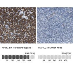
- Experimental details
- Immunohistochemistry analysis in human parathyroid gland and lymph node tissues using Anti-MARC2 antibody. Corresponding MARC2 RNA-seq data are presented for the same tissues.
- Sample type
- HUMAN
Enhanced validation
- Submitted by
- Atlas Antibodies (provider)
- Enhanced method
- Independent antibody validation
- Main image
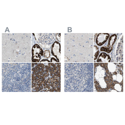
- Experimental details
- Immunohistochemical staining of human cerebral cortex, kidney, lymph node and parathyroid gland using Anti-MARC2 antibody HPA015085 (A) shows similar protein distribution across tissues to independent antibody HPA017572 (B).

Supportive validation
- Submitted by
- Atlas Antibodies (provider)
- Main image
- Experimental details
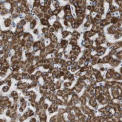
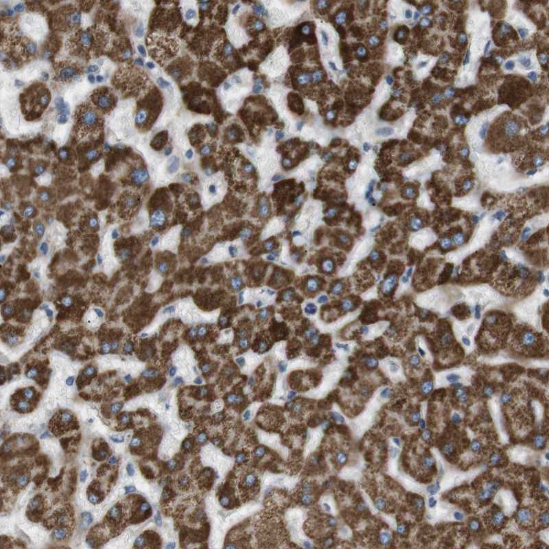
- Immunohistochemical staining of human liver shows strong cytoplasmic positivity in hepatocytes.
- Submitted by
- Atlas Antibodies (provider)
- Main image
- Experimental details
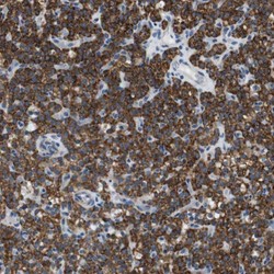
- Immunohistochemical staining of human parathyroid gland shows high expression.
- Sample type
- HUMAN

- Submitted by
- Atlas Antibodies (provider)
- Main image
- Experimental details
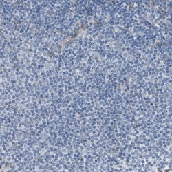
- Immunohistochemical staining of human lymph node shows low expression as expected.
- Sample type
- HUMAN

- Submitted by
- Atlas Antibodies (provider)
- Main image
- Experimental details
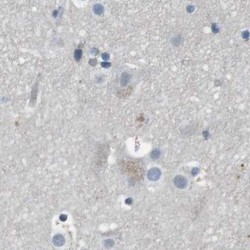
- Immunohistochemical staining of human cerebral cortex using Anti-MARC2 antibody HPA015085.
- Sample type
- HUMAN

- Submitted by
- Atlas Antibodies (provider)
- Main image
- Experimental details
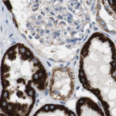
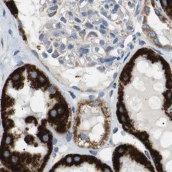
- Immunohistochemical staining of human kidney using Anti-MARC2 antibody HPA015085.
- Sample type
- HUMAN