Explore
 Validate
 Learn
LearnNBP2-15791
antibody from Novus Biologicals
Targeting: CD274
B7-H, B7-H1, B7H1, PD-L1, PDCD1LG1, PDL1
 Western blot
 Immunocytochemistry
Antibody data
- Antibody Data
- Antigen structure
- References [0]
- Comments [0]
- Validations
- Western blot [8]
- Immunohistochemistry [3]
Submit
Validation data
Reference
Comment
Report error
- Product number
- NBP2-15791 - Provider product page

- Provider
- Novus Biologicals
- Product name
- Rabbit Polyclonal PD-L1 Antibody
- Antibody type
- Polyclonal
- Description
- Immunogen affinity purified.
- Reactivity
- Human
- Host
- Rabbit
- Isotype
- IgG
- Vial size
- 0.1 ml
- Storage
- Aliquot and store at -20C or -80C. Avoid freeze-thaw cycles.
No comments: Submit comment
Supportive validation
- Submitted by
- Novus Biologicals (provider)
- Main image
- Experimental details
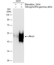
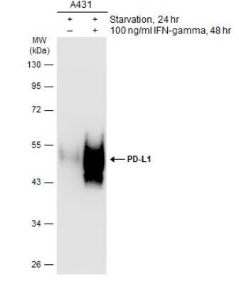
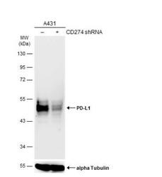
- Western Blot: PD-L1 Antibody [NBP2-15791] - PD-L1/B7-H1 Antibody [NBP2-15791] - Untreated (-) and treated (+) A431 whole cell extracts (30 ug) were separated by 10% SDS-PAGE, and the membranes were blotted with PD-L1 antibody. The HRP-conjugated anti-rabbit IgG antibody (NBP2-19301) was used to detect the primary antibody.
- Submitted by
- Novus Biologicals (provider)
- Main image
- Experimental details
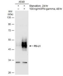
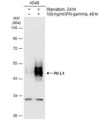
- Western Blot: PD-L1 Antibody [NBP2-15791] - PD-L1/B7-H1 Antibody [NBP2-15791] - Untreated (-) and treated (+) A549 whole cell extracts (30 ug) were separated by 10% SDS-PAGE, and the membranes were blotted with PD-L1 antibody. The HRP-conjugated anti-rabbit IgG antibody (NBP2-19301) was used to detect the primary antibody.
- Submitted by
- Novus Biologicals (provider)
- Main image
- Experimental details
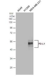
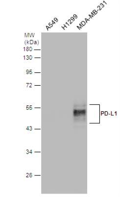
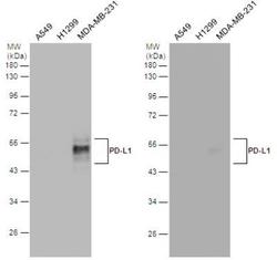
- Western Blot: PD-L1/B7-H1 Antibody [NBP2-15791] - Various whole cell extracts (30 ug) were separated by 12% SDS-PAGE, and the membranes were blotted with PD-L1 antibody diluted at 1:2000. The HRP-conjugated anti-rabbit IgG antibody (NBP2-19301) was used to detect the primary antibody.
- Submitted by
- Novus Biologicals (provider)
- Main image
- Experimental details
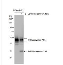
- Western Blot: PD-L1/B7-H1 Antibody [NBP2-15791] - Untreated (-) and treated (+) MDA-MB-231 whole cell extracts (30 ug) were separated by 10% SDS-PAGE, and the membrane was blotted with PD-L1 antibody.

- Submitted by
- Novus Biologicals (provider)
- Main image
- Experimental details
- Western Blot: PD-L1 Antibody [NBP2-15791] - PD-L1/B7-H1 Antibody [NBP2-15791] - Non-transfected (-) and transfected (+) A431 whole cell extracts (30 ug) were separated by 10% SDS-PAGE, and the membrane was blotted with PD-L1 antibody. HRP-conjugated anti-rabbit IgG antibody (NBP2-19301) was used to detect the primary antibody.

- Submitted by
- Novus Biologicals (provider)
- Main image
- Experimental details
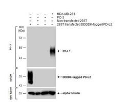
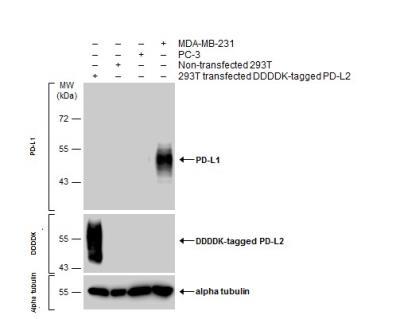
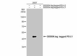
- Western Blot: PD-L1/B7-H1 Antibody [NBP2-15791] - Various whole cell extracts were separated by 10% SDS-PAGE, and the membranes were blotted with PD-L1 antibody diluted at 1:600 and with DDDDK tag antibody (NBP2-43574) diluted at 1:3000 to detect DDDDK-tagged PD-L2. The HRP-conjugated anti-rabbit IgG antibody (NBP2-19301) was used to detect the primary antibody.
- Submitted by
- Novus Biologicals (provider)
- Main image
- Experimental details
- Western Blot: PD-L1 Antibody [NBP2-15791] - PD-L1/B7-H1 Antibody [NBP2-15791] - Various whole cell extracts (30 ug) were separated by 12% SDS-PAGE, and the membranes were blotted with PD-L1 antibody. HRP-conjugated anti-rabbit IgG antibody was used to detect the primary antibody. NBP2-15791 on the left and competitor's antibody on the right.

- Submitted by
- Novus Biologicals (provider)
- Main image
- Experimental details
- Western Blot: PD-L1 Antibody [NBP2-15791] - Non-transfected (-) and transfected (+) 293T whole cell extracts (30 ug) were separated by 10% SDS-PAGE, and the membrane was blotted with PD-L1 antibody diluted at 1:1000. The HRP-conjugated anti-rabbit IgG antibody (NBP2-19301) was used to detect the primary antibody, and the signal was developed with Trident ECL plus-Enhanced.

Supportive validation
- Submitted by
- Novus Biologicals (provider)
- Main image
- Experimental details
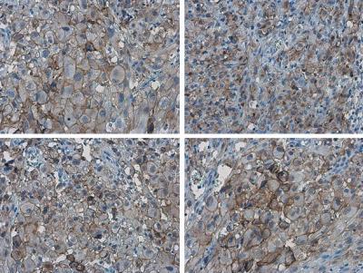
- Immunohistochemistry-Paraffin: PD-L1/B7-H1 Antibody [NBP2-15791] - PD-L1 proteinat cell membrane in human ovarian carcinoma by immunohistochemical analysis. Sample: Paraffin-embedded human ovarian carcinoma. PD-L1 antibody diluted at 1:1000.
- Submitted by
- Novus Biologicals (provider)
- Main image
- Experimental details
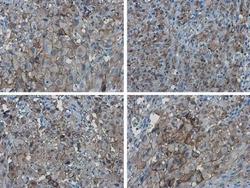
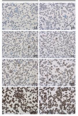
- Immunohistochemistry-Paraffin: PD-L1 Antibody [NBP2-15791] - Detection of cell membranes in PD-L1 protein-expressing cell lines by immunohistochemical analysis. Antibodies: PD-L1 antibody, and competitor's antibody. Samples: Negative (-), low positive (+), intermediate positive (++) and strong positive (+++) cell line cores assessed using Quantitative Digital Pathology.
- Submitted by
- Novus Biologicals (provider)
- Main image
- Experimental details
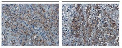
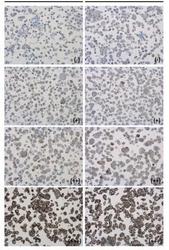
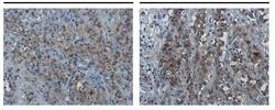
- Immunohistochemistry-Paraffin: PD-L1/B7-H1 Antibody [NBP2-15791] - PD-L1 antibody detects PD-L1 protein at cell membrane in human ovarian carcinoma by immunohistochemical analysis. Antibodies: PD-L1 antibody, and competitor's antibody.