 Explore
 Validate
 Learn
Learn Western blot
 Immunocytochemistry
Antibody data
- Antibody Data
- Antigen structure
- References [2]
- Comments [0]
- Validations
- Western blot [1]
- Immunohistochemistry [5]
- Flow cytometry [2]
Submit
Validation data
Reference
Comment
Report error
- Product number
- NBP2-36571 - Provider product page

- Provider
- Novus Biologicals
- Product name
- Mouse Monoclonal Calnexin Antibody
- Antibody type
- Monoclonal
- Description
- Protein G purified.
- Reactivity
- Human
- Host
- Mouse
- Isotype
- IgG
- Vial size
- 0.1 mg
- Concentration
- 1.0 mg/ml
- Storage
- Store at 4C short term. Aliquot and store at -20C long term. Avoid freeze-thaw cycles.
Submitted references Exosomal miR-16-5p as a target for malignant mesothelioma.
Actin polymerization plays a significant role in asbestos-induced inflammasome activation in mesothelial cells in vitro.
Munson PB, Hall EM, Farina NH, Pass HI, Shukla A
Scientific reports 2019 Aug 12;9(1):11688
Scientific reports 2019 Aug 12;9(1):11688
Actin polymerization plays a significant role in asbestos-induced inflammasome activation in mesothelial cells in vitro.
MacPherson M, Westbom C, Kogan H, Shukla A
Histochemistry and cell biology 2017 May;147(5):595-604
Histochemistry and cell biology 2017 May;147(5):595-604
No comments: Submit comment
Supportive validation
- Submitted by
- Novus Biologicals (provider)
- Main image
- Experimental details
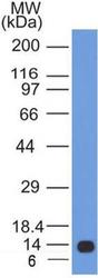
- Western Blot: Calnexin Antibody (IE2.1C12) [NBP2-36571] - Analysis of 11 kDa Partial Recombinant Human Calnexin protein with Calnexin antibody (clone IE2.1C12) at 0.5 ug/ml concentration.

Supportive validation
- Submitted by
- Novus Biologicals (provider)
- Main image
- Experimental details
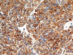
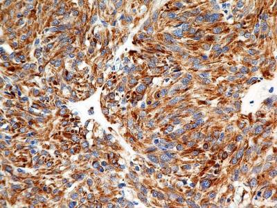
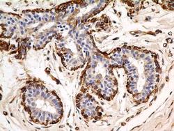
- Immunohistochemistry-Paraffin: Calnexin Antibody (IE2.1C12) [NBP2-36571] - Analysis of FFPE tissue section of malignant stromal tumor of the human small bowel using mouse monoclonal Calnexin antibody (clone IE2.1C12) at 7 ug/ml concentration. The cancer cells showed a very strong cytoplasmic reactivity for Calnexin.
- Submitted by
- Novus Biologicals (provider)
- Main image
- Experimental details
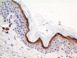
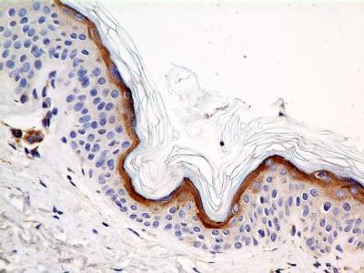
- Immunohistochemistry-Paraffin: Calnexin Antibody (IE2.1C12) [NBP2-36571] - Analysis of FFPE tissue section of human skin using mouse monoclonal Calnexin antibody (clone IE2.1C12) at 7 ug/ml concentration. The outermost keratinocytes layer of the epidermis showed cytoplasmic positivity for Calnexin protein.
- Submitted by
- Novus Biologicals (provider)
- Main image
- Experimental details
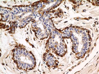
- Immunohistochemistry-Paraffin: Calnexin Antibody (IE2.1C12) [NBP2-36571] - Analysis of FFPE tissue section of normal human breast using mouse monoclonal Calnexin antibody (clone IE2.1C12) at 7 ug/ml concentration. The myoepithelial cells around the lobules depicted a very strong cytoplasmic staining.
- Submitted by
- Novus Biologicals (provider)
- Main image
- Experimental details
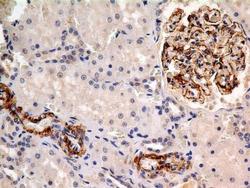
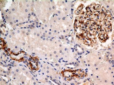
- Immunohistochemistry-Paraffin: Calnexin Antibody (IE2.1C12) [NBP2-36571] - Analysis of FFPE tissue section of normal human kidney using mouse monoclonal Calnexin antibody (clone IE2.1C12) at 7 ug/ml concentration. The cells of Glomeruli developed strong cytoplasmic staining.
- Submitted by
- Novus Biologicals (provider)
- Main image
- Experimental details
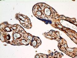
- Immunohistochemistry-Paraffin: Calnexin Antibody (IE2.1C12) [NBP2-36571] - Analysis of FFPE tissue section of human placenta using mouse monoclonal Calnexin antibody (clone IE2.1C12) at 7 ug/ml concentration.

Supportive validation
- Submitted by
- Novus Biologicals (provider)
- Main image
- Experimental details
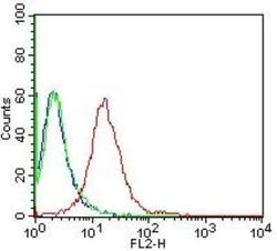
- Flow (Intracellular): Calnexin Antibody (IE2.1C12) [NBP2-36571] - Intracellular staining of human Calnexin in Flow cytometry using 5.0 ug of antibody per 1 million cells. Isotype control was mouse IgG2b kappa.
- Submitted by
- Novus Biologicals (provider)
- Main image

- Experimental details
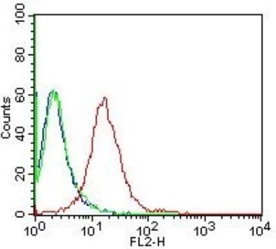
- Flow Cytometry: Calnexin Antibody (IE2.1C12) [NBP2-36571] - Analysis of Allophycocyanin conjugate of NBP2-36571. An intracellular stain was performed on Jurkat cells with Calnexin antibody (IE2.1C12) NBP2-36571APC (blue) and a matched isotype control (orange). Cells were fixed with 4% PFA and then permeablized with 0.1% saponin.
