 Explore
 Validate
 Learn
Learn Western blot
 Immunohistochemistry
Antibody data
- Antibody Data
- Antigen structure
- References [2]
- Comments [0]
- Validations
- Immunohistochemistry [1]
Submit
Validation data
Reference
Comment
Report error
- Product number
- MAB475-050 - Provider product page

- Provider
- R&D Systems
- Product name
- Mouse Wnt-4 Antibody
- Antibody type
- Monoclonal
- Description
- Protein A or G purified from hybridoma culture supernatant. Detects mouse Wnt-4 in direct ELISAs and Western blots.
- Reactivity
- Mouse
- Host
- Rat
- Conjugate
- Unconjugated
- Antigen sequence
P22724- Isotype
- IgG
- Antibody clone number
- 55010
- Vial size
- 50 ug
- Concentration
- LYOPH
- Storage
- Use a manual defrost freezer and avoid repeated freeze-thaw cycles. 12 months from date of receipt, -20 to -70 °C as supplied. 1 month, 2 to 8 °C under sterile conditions after reconstitution. 6 months, -20 to -70 °C under sterile conditions after reconstitution.
Submitted references Macrophages promote epithelial proliferation following infectious and non-infectious lung injury through a Trefoil factor 2-dependent mechanism.
Wnt glycoproteins regulate the expression of FoxN1, the gene defective in nude mice.
Hung LY, Sen D, Oniskey TK, Katzen J, Cohen NA, Vaughan AE, Nieves W, Urisman A, Beers MF, Krummel MF, Herbert DR
Mucosal immunology 2019 Jan;12(1):64-76
Mucosal immunology 2019 Jan;12(1):64-76
Wnt glycoproteins regulate the expression of FoxN1, the gene defective in nude mice.
Balciunaite G, Keller MP, Balciunaite E, Piali L, Zuklys S, Mathieu YD, Gill J, Boyd R, Sussman DJ, Holländer GA
Nature immunology 2002 Nov;3(11):1102-8
Nature immunology 2002 Nov;3(11):1102-8
No comments: Submit comment
Supportive validation
- Submitted by
- R&D Systems (provider)
- Main image
- Experimental details
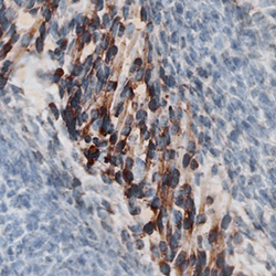
- Wnt-4 in Mouse Embryo. Wnt-4 was detected in immersion fixed frozen sections of mouse embryo (13 d.p.c.) using Rat Anti-Mouse Wnt-4 Monoclonal Antibody (Catalog # MAB475) at 25 µg/mL overnight at 4 °C. Tissue was stained using the Anti-Rat HRP-DAB Cell & Tissue Staining Kit (brown; Catalog # CTS017) and counterstained with hematoxylin (blue). Specific staining was localized to muscle cells. View our protocol for Chromogenic IHC Staining of Frozen Tissue Sections.
