 Explore
 Validate
 Learn
Learn Western blot
Antibody data
- Antibody Data
- Antigen structure
- References [0]
- Comments [0]
- Validations
- Western blot [6]
- Immunocytochemistry [2]
- Immunohistochemistry [1]
Submit
Validation data
Reference
Comment
Report error
- Product number
- PA5-29159 - Provider product page

- Provider
- Invitrogen Antibodies
- Product name
- BID Polyclonal Antibody
- Antibody type
- Polyclonal
- Antigen
- Recombinant full-length protein
- Description
- Recommended positive controls: 293T, A431, H1299, HeLa, HepG2, Molt-4, Raji.
- Concentration
- 1 mg/mL
No comments: Submit comment
Supportive validation
- Submitted by
- Invitrogen Antibodies (provider)
- Main image
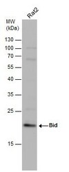
- Experimental details
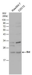
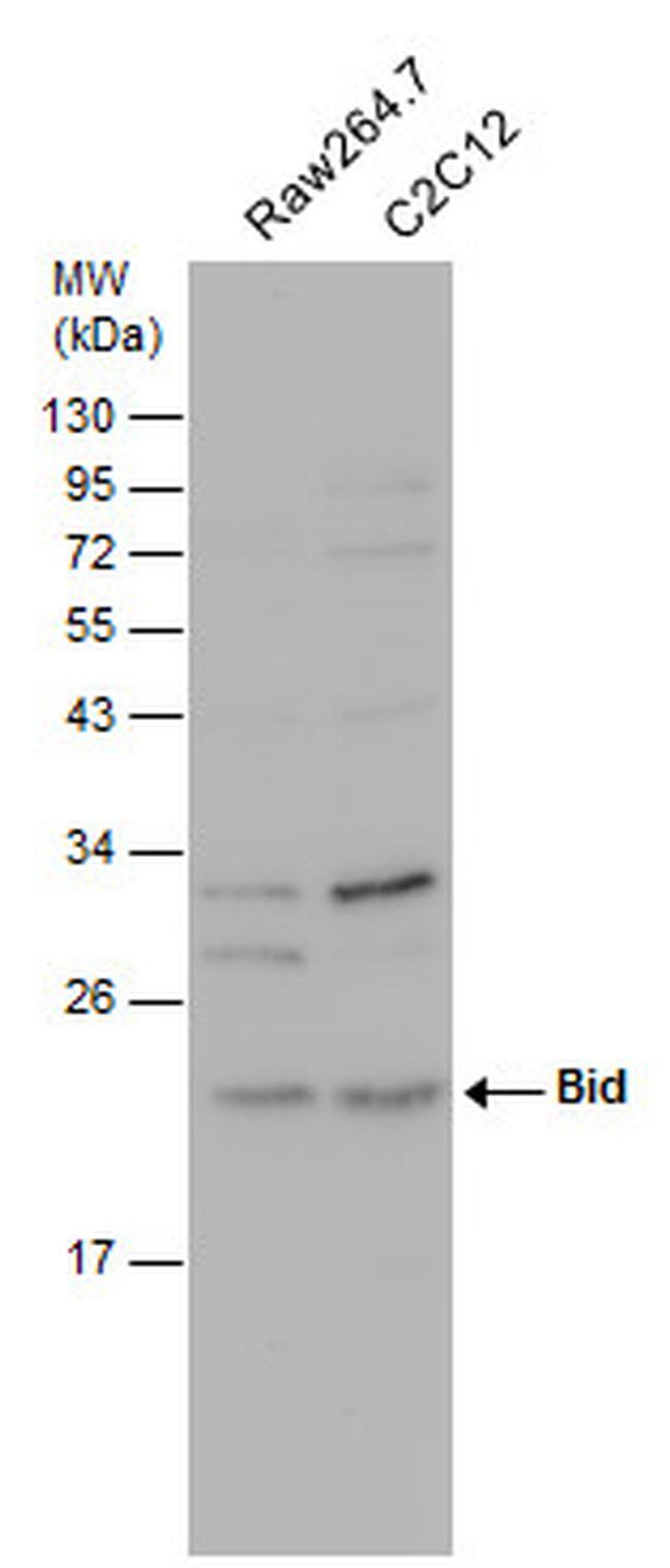
- Western blot analysis of Bid in Various whole cell extracts (30 µg). Samples were separated by 12% SDS-PAGE and the membrane was probed with Bid Polyclonal antibody (Product # PA5-29159) at a dilution of 1:500.
- Submitted by
- Invitrogen Antibodies (provider)
- Main image
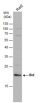
- Experimental details
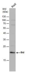
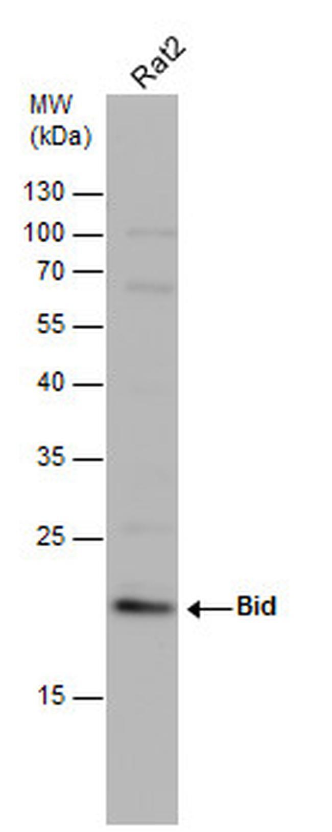
- Western blot analysis of Bid in whole cell extract (30 µg). Samples was separated by 12% SDS-PAGE and the membrane was probed with Bid Polyclonal antibody (Product # PA5-29159) at a dilution of 1:1000.
- Submitted by
- Invitrogen Antibodies (provider)
- Main image
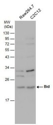
- Experimental details
- Western Blot using BID Polyclonal Antibody (Product # PA5-29159). Whole cell extract (30 µg) was separated by 12% SDS-PAGE, and the membrane was blotted with BID Polyclonal Antibody (Product # PA5-29159) diluted at 1:1,000.
- Submitted by
- Invitrogen Antibodies (provider)
- Main image
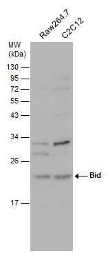
- Experimental details
- Western Blot using BID Polyclonal Antibody (Product # PA5-29159). Various whole cell extracts (30 µg) were separated by 12% SDS-PAGE, and the membrane was blotted with BID Polyclonal Antibody (Product # PA5-29159) diluted at 1:500.
- Submitted by
- Invitrogen Antibodies (provider)
- Main image
- Experimental details
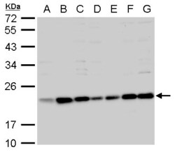
- BID Polyclonal Antibody detects Bid protein by western blot analysis. A. 30 µg 293T whole cell lysate/extract. B. 30 µg A431 whole cell lysate/extract. C. 30 µg H1299 whole cell lysate/extract. D. 30 µg HeLa whole cell lysate/extract. E. 30 µg HepG2 whole cell lysate/extract. F. 30 µg Molt-4 whole cell lysate/extract. G. 30 µg Raji whole cell lysate/extract.7.5 % SDS-PAGE. BID Polyclonal Antibody (Product # PA5-29159) dilution: 1:1,000.

- Submitted by
- Invitrogen Antibodies (provider)
- Main image
- Experimental details
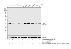
- Western blot was performed using Anti-BID Polyclonal Antibody (Product # PA5-29159) and a 20 kDa band corresponding to BID was observed across cell lines and decreased upon Staurosporine, Etoposide and also with Actinomycin D along with TNF Alpha treatment. Whole cell extracts (30 µg lysate) of A-431 (Lane 1), A-431 treated with Staurosporine (1uM for 4 hr) (Lane 2), A-431 treated with Staurosporine (1uM for 8 hr) (Lane 3), A-431 treated with Etoposide (25uM for 8 hr) (Lane 4), A-431 treated with Actinomycin along with TNF Alpha (5ug/ml and 10ng/ml for 5 hr) (Lane 5), MCF-7 (Lane 6), Jurkat (Lane 7), MOLT-4 (Lane 8), PC-3 (Lane 9) and SH-SY5Y (Lane 10) were electrophoresed using Novex® NuPAGE® 12 % Bis-Tris gel (Product # NP0342BOX). Resolved proteins were then transferred onto a nitrocellulose membrane (Product # IB23001) by iBlot® 2 Dry Blotting System (Product # IB21001). The blot was probed with the primary antibody (1:3000 dilution) and detected by chemiluminescence with Goat anti-Rabbit IgG (H+L) Superclonal™ Recombinant Secondary Antibody, HRP (Product # A27036, 1:4000 dilution) using the iBright FL 1000 (Product # A32752). Chemiluminescent detection was performed using Novex® ECL Chemiluminescent Substrate Reagent Kit (Product # WP20005).

Supportive validation
- Submitted by
- Invitrogen Antibodies (provider)
- Main image
- Experimental details
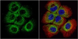
- Immunocytochemistry-Immunofluorescence analysis of BID was performed in A431 cells fixed in 4% paraformaldehyde at RT for 15 min. Green: BID Polyclonal Antibody (Product # PA5-29159) diluted at 1:1000. Red: alpha Tubulin, a cytoskeleton marker. Blue: Hoechst 33342 staining.

- Submitted by
- Invitrogen Antibodies (provider)
- Main image
- Experimental details
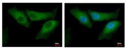
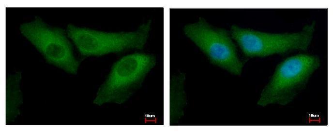
- BID Polyclonal Antibody detects BID protein at cytoplasm by immunofluorescent analysis. Sample: HeLa cells were fixed in 2% paraformaldehyde/culture medium at 37ºC for 30 min. Green: BID protein stained by BID Polyclonal Antibody (Product # PA5-29159) diluted at 1:500. Blue: Hoechst 33343 staining.
Supportive validation
- Submitted by
- Invitrogen Antibodies (provider)
- Main image
- Experimental details
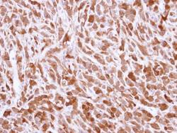
- Immunohistochemical analysis of paraffin-embedded U87 xenograft, using Bid (Product # PA5-29159) antibody at 1:500 dilution. Antigen Retrieval: Citrate buffer, pH 6.0, 15 min.
