Explore
 Validate
 Learn
Learn Western blot
 Flow cytometry
Antibody data
- Antibody Data
- Antigen structure
- References [0]
- Comments [0]
- Validations
- Western blot [3]
- Immunohistochemistry [9]
Submit
Validation data
Reference
Comment
Report error
- Product number
- MA5-25459 - Provider product page

- Provider
- Invitrogen Antibodies
- Product name
- NDUFB9 Monoclonal Antibody (OTI13H11)
- Antibody type
- Monoclonal
- Antigen
- Recombinant protein fragment
- Reactivity
- Human
- Host
- Mouse
- Isotype
- IgG
- Antibody clone number
- OTI13H11
- Vial size
- 100 µL
- Concentration
- 1 mg/mL
- Storage
- -20° C, Avoid Freeze/Thaw Cycles
No comments: Submit comment
Supportive validation
- Submitted by
- Invitrogen Antibodies (provider)
- Main image
- Experimental details
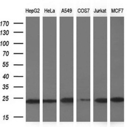
- Western blot analysis of NDUFB9 in HepG2, HeLa, A549, COS7, Jurkat, MCF7 cells using 10 µg per lane. Samples were probed with NDUFB9 (Product # MA5-25459) monoclonal antibody at a dilution of 1:200.

- Submitted by
- Invitrogen Antibodies (provider)
- Main image
- Experimental details
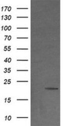
- Western blot analysis of NDUFB9 in HEK293T cells in untransfected (Left lane) and transfected (Right lane) samples using 5 µg per lane. The samples were separated by SDS-PAGE and probed with NDUFB9 (Product # MA5-25459) monoclonal antibody.

- Submitted by
- Invitrogen Antibodies (provider)
- Main image
- Experimental details
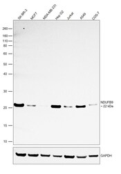
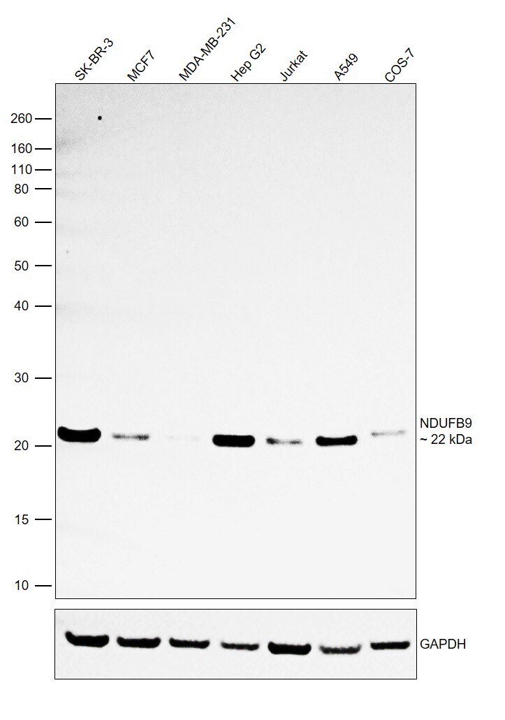
- Western Blot was performed using Anti-NDUFB9 Monoclonal Antibody (OTI13H11) (Product # MA5-25459) and a 22 kDa band corresponding to NADH dehydrogenase [ubiquinone] 1 beta subcomplex subunit 9 was observed. Whole cell extracts (30 µg lysate) of SK-BR-3 (Lane 1), MCF7 (Lane 2), MDA-MB-231 (Lane 3), Hep G2 (Lane 4), Jurkat (Lane 5), A549 (Lane 6) and COS-7 (Lane 7) were electrophoresed using NuPAGE™ 10% Bis-Tris Protein Gel (Product # NP0302BOX). Resolved proteins were then transferred onto a nitrocellulose membrane (Product # IB23001) by iBlot® 2 Dry Blotting System (Product # IB21001). The blot was probed with the primary antibody (1:500) and detected by chemiluminescence with Goat anti-Mouse IgG (H+L) Superclonal™ Recombinant Secondary Antibody, HRP (Product # A28177, 1:4000) using the iBright FL 1000 (Product # A32752). Chemiluminescent detection was performed using Novex® ECL Chemiluminescent Substrate Reagent Kit (Product # WP20005). Relative expression was observed between SK-BR-3, MCF7 and MDA-MB-231 as expected (https://doi.org/10.1371/journal.pone.0144441).
Supportive validation
- Submitted by
- Invitrogen Antibodies (provider)
- Main image
- Experimental details
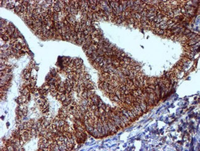
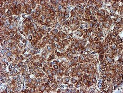
- Immunohistochemistry was performed on paraffin-embedded carcinoma of human liver tissue. To expose target proteins, 10mM citric buffer, pH6.0, 120°C for 3min was used. Following antigen retrieval, tissues were probed with a NDUFB9 monoclonal antibody (Product # MA5-25459).

- Submitted by
- Invitrogen Antibodies (provider)
- Main image

- Experimental details
- Immunohistochemistry was performed on paraffin-embedded adenocarcinoma of human colon tissue. To expose target proteins, 10mM citric buffer, pH6.0, 120°C for 3min was used. Following antigen retrieval, tissues were probed with a NDUFB9 monoclonal antibody (Product # MA5-25459).

- Submitted by
- Invitrogen Antibodies (provider)
- Main image

- Experimental details
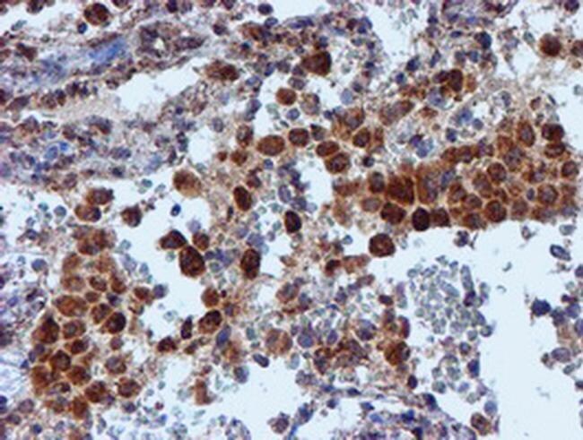
- Immunohistochemistry was performed on paraffin-embedded carcinoma of human lung tissue. To expose target proteins, 10mM citric buffer, pH6.0, 120°C for 3min was used. Following antigen retrieval, tissues were probed with a NDUFB9 monoclonal antibody (Product # MA5-25459).
- Submitted by
- Invitrogen Antibodies (provider)
- Main image

- Experimental details
- Immunohistochemistry was performed on paraffin-embedded human liver tissue. To expose target proteins, 10mM citric buffer, pH6.0, 120°C for 3min was used. Following antigen retrieval, tissues were probed with a NDUFB9 monoclonal antibody (Product # MA5-25459).

- Submitted by
- Invitrogen Antibodies (provider)
- Main image
- Experimental details
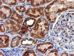
- Immunohistochemistry was performed on paraffin-embedded human kidney tissue. To expose target proteins, 10mM citric buffer, pH6.0, 120°C for 3min was used. Following antigen retrieval, tissues were probed with a NDUFB9 monoclonal antibody (Product # MA5-25459).

- Submitted by
- Invitrogen Antibodies (provider)
- Main image
- Experimental details
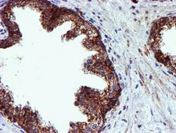
- Immunohistochemistry was performed on paraffin-embedded carcinoma of human prostate tissue. To expose target proteins, 10mM citric buffer, pH6.0, 120°C for 3min was used. Following antigen retrieval, tissues were probed with a NDUFB9 monoclonal antibody (Product # MA5-25459).

- Submitted by
- Invitrogen Antibodies (provider)
- Main image
- Experimental details
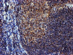
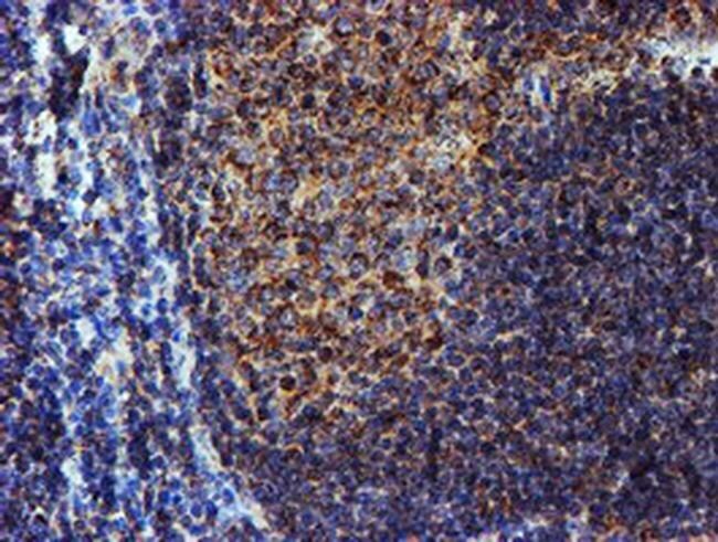
- Immunohistochemistry was performed on paraffin-embedded human tonsil tissue. To expose target proteins, 10mM citric buffer, pH6.0, 120°C for 3min was used. Following antigen retrieval, tissues were probed with a NDUFB9 monoclonal antibody (Product # MA5-25459).
- Submitted by
- Invitrogen Antibodies (provider)
- Main image

- Experimental details
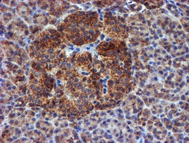
- Immunohistochemistry was performed on paraffin-embedded human pancreas tissue. To expose target proteins, 10mM citric buffer, pH6.0, 120°C for 3min was used. Following antigen retrieval, tissues were probed with a NDUFB9 monoclonal antibody (Product # MA5-25459).
- Submitted by
- Invitrogen Antibodies (provider)
- Main image

- Experimental details
- Immunohistochemistry was performed on paraffin-embedded adenocarcinoma of human endometrium tissue. To expose target proteins, 10mM citric buffer, pH6.0, 120°C for 3min was used. Following antigen retrieval, tissues were probed with a NDUFB9 monoclonal antibody (Product # MA5-25459).