 Explore
 Validate
 Learn
Learn Western blot
Antibody data
- Antibody Data
- Antigen structure
- References [1]
- Comments [0]
- Validations
- Western blot [3]
Submit
Validation data
Reference
Comment
Report error
- Product number
- AF13662 - Provider product page

- Provider
- R&D Systems
- Product name
- Human PTP1B Antibody
- Antibody type
- Polyclonal
- Description
- Antigen Affinity-purified. Detects endogenous human PTP1B and does not cross-react with recombinant mouse PTP1B in Western blots.
- Reactivity
- Human
- Host
- Goat
- Conjugate
- Unconjugated
- Antigen sequence
P18031- Isotype
- IgG
- Vial size
- 100 ug
- Concentration
- LYOPH
- Storage
- Use a manual defrost freezer and avoid repeated freeze-thaw cycles. 12 months from date of receipt, -20 to -70 °C as supplied. 1 month, 2 to 8 °C under sterile conditions after reconstitution. 6 months, -20 to -70 °C under sterile conditions after reconstitution.
Submitted references Absence of leptin resistance in platelets from morbidly obese individuals may contribute to the increased thrombosis risk in obesity.
Dellas C, Schäfer K, Rohm I, Lankeit M, Ellrott T, Faustin V, Riggert J, Hasenfuss G, Konstantinides S
Thrombosis and haemostasis 2008 Dec;100(6):1123-9
Thrombosis and haemostasis 2008 Dec;100(6):1123-9
No comments: Submit comment
Supportive validation
- Submitted by
- R&D Systems (provider)
- Main image

- Experimental details
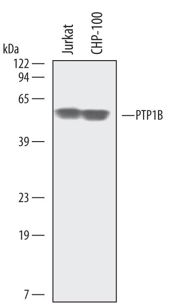
- Detection of Human PTP1B by Western Blot. Western blot shows lysates of Jurkat human acute T cell leukemia cell line and CHP-100 human neuroblastoma cell line. PVDF membrane was probed with 0.3 µg/mL of Human PTP1B Antigen Affinity-purified Polyclonal Antibody (Catalog # AF13662) followed by HRP-conjugated Anti-Goat IgG Secondary Antibody (Catalog # HAF109). A specific band was detected for PTP1B at approximately 50 kDa (as indicated). This experiment was conducted under reducing conditions and using Immunoblot Buffer Group 1.
- Submitted by
- R&D Systems (provider)
- Main image

- Experimental details
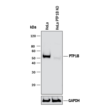
- Western Blot Shows Human PTP1B Specificity by Using Knockout Cell Line. Western blot shows lysates of HeLa human cervical epithelial carcinoma parental cell line and PTP1B knockout HeLa cell line (KO). PVDF membrane was probed with 0.3 µg/mL of Goat Anti-Human PTP1B Antigen Affinity-purified Polyclonal Antibody (Catalog # AF13662) followed by HRP-conjugated Anti-Goat IgG Secondary Antibody (Catalog # HAF017). A specific band was detected for PTP1B at approximately 52 kDa (as indicated) in the parental HeLa cell line, but is not detectable in knockout HeLa cell line. GAPDH (Catalog # AF5718) is shown as a loading control. This experiment was conducted under reducing conditions and using Immunoblot Buffer Group 1.
- Submitted by
- R&D Systems (provider)
- Main image
- Experimental details
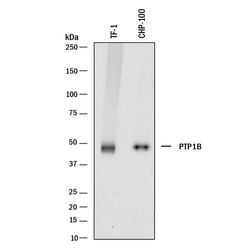
- Detection of Human PTP1B by Western Blot. Western blot shows lysates of TF-1 human erythroleukemic cell line and CHP-100 human neuroblastoma cell line. PVDF membrane was probed with 0.05 µg/mL of Goat Anti-Human PTP1B Antigen Affinity-purified Polyclonal Antibody (Catalog # AF13662) followed by HRP-conjugated Anti-Goat IgG Secondary Antibody (Catalog # HAF017). A specific band was detected for PTP1B at approximately 50 kDa (as indicated). This experiment was conducted under reducing conditions and using Immunoblot Buffer Group 1.
