Explore
 Validate
 Learn
Learn Western blot
Antibody data
- Antibody Data
- Antigen structure
- References [0]
- Comments [0]
- Validations
- Western blot [3]
- Immunocytochemistry [4]
Submit
Validation data
Reference
Comment
Report error
- Product number
- PA5-34433 - Provider product page

- Provider
- Invitrogen Antibodies
- Product name
- DLK1 Polyclonal Antibody
- Antibody type
- Polyclonal
- Antigen
- Synthetic peptide
- Description
- A suggested positive control is HepG2 cell lysate.
- Concentration
- 1 mg/mL
No comments: Submit comment
Supportive validation
- Submitted by
- Invitrogen Antibodies (provider)
- Main image
- Experimental details
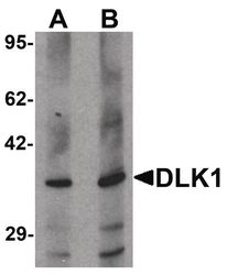
- Western blot analysis of 293 cell lysate using a DLK1 polyclonal antibody (Product # PA5-34433) at (A) 1 and (B) 2 µg/mL.
- Submitted by
- Invitrogen Antibodies (provider)
- Main image

- Experimental details
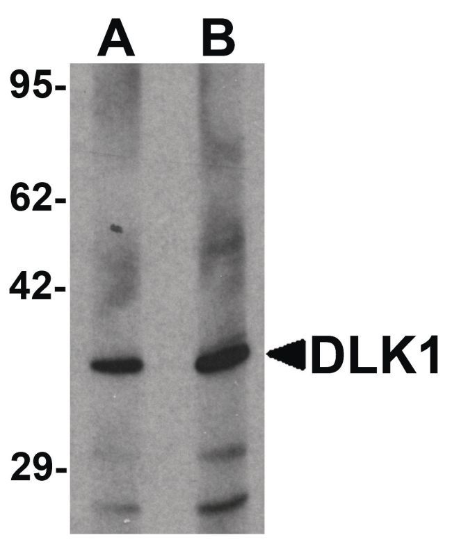
- Western Blot analysis of HepG2 in 293 cell lysate with DLK1 Polyclonal Antibody (Product # PA5-34433) at (A) 1 and (B) 2 µg/mL.

- Submitted by
- Invitrogen Antibodies (provider)
- Main image
- Experimental details
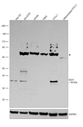
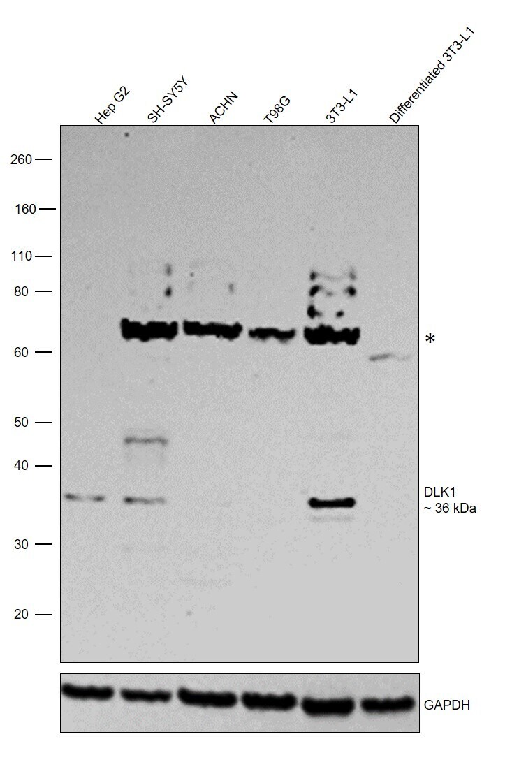
- Western blot was performed using Anti-DLK1 Polyclonal Antibody (Product # PA5-34433) and a 36 kDa band corresponding to Protein delta homolog 1 was observed along with a strong uncharacterized band (*) around 70 kDa. Whole cell extracts (30 µg lysate) of Hep G2 (Lane 1), SH-SY5Y (Lane 2), ACHN (Lane 3), T98G (Lane 4), 3T3-L1 (Lane 5) and 3T3-L1 differentiated to adipocytes (Lane 6) were electrophoresed using NuPAGE™ 4-12% Bis-Tris Protein Gel (Product # NP0322BOX). Resolved proteins were then transferred onto a nitrocellulose membrane (Product # IB23001) by iBlot® 2 Dry Blotting System (Product # IB21001). The blot was probed with the primary antibody (1 µg/mL ) and detected by chemiluminescence with Goat anti-Rabbit IgG (H+L) Superclonal™ Recombinant Secondary Antibody, HRP (Product # A27036,1:20,000) using the iBright FL 1000 (Product # A32752). Chemiluminescent detection was performed using SuperSignal™ West Pico PLUS Chemiluminescent Substrate (Product # 34580). Band of interest was prominent in 3T3-L1, whereas it was absent in 3T3-L1 differentiated to adipocytes as expected (DOI 10.1074/jbc.M500463200). Expression was found to be null in ACHN (https://doi.org/10.1093/hmg/ddl001) and T98G (https://doi.org/10.1038/sj.onc.1209219) as well in accordance with the reports.
Supportive validation
- Submitted by
- Invitrogen Antibodies (provider)
- Main image
- Experimental details
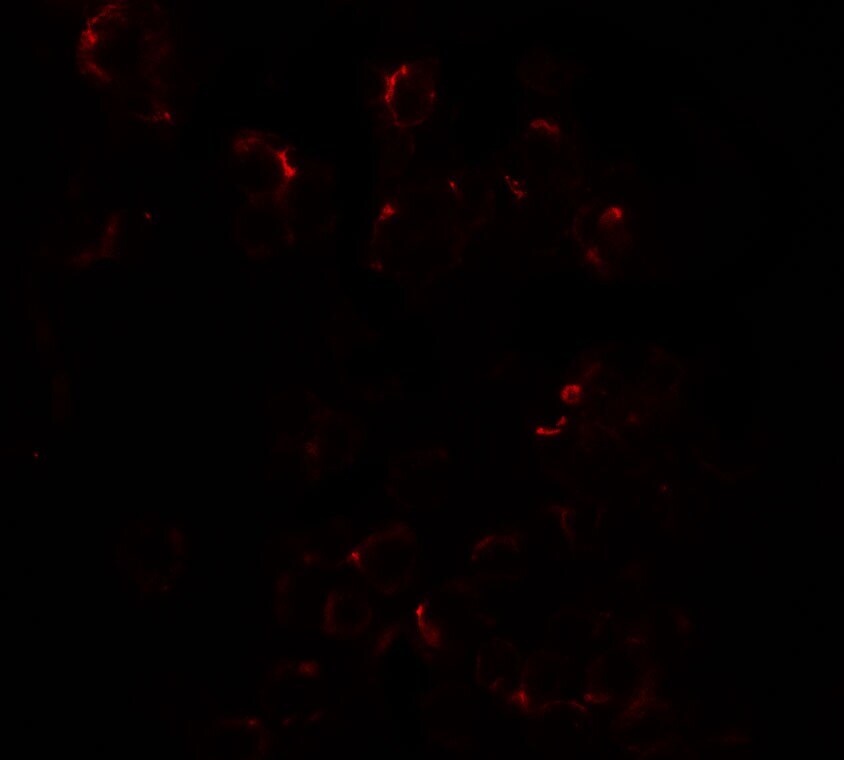
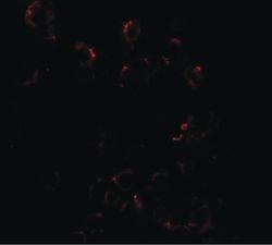
- Immunofluorescent analysis of HepG2 cells using a DLK1 polyclonal antibody (Product # PA5-34433) at a 20 µg/mL dilution.

- Submitted by
- Invitrogen Antibodies (provider)
- Main image

- Experimental details
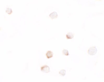
- Immunocytochemistry staining of HepG2 cells using a DLK1 polyclonal antibody (Product # PA5-34433) at a 25 µg/mL dilution.

- Submitted by
- Invitrogen Antibodies (provider)
- Main image

- Experimental details
- Immunocytochemistry of DLK1 in HepG2 cells with DLK1 Polyclonal Antibody (Product # PA5-34433) at 2.5 µg/mL.
- Submitted by
- Invitrogen Antibodies (provider)
- Main image

- Experimental details
- Immunofluorescence of DLK1 in HepG2 cells with DLK1 Polyclonal Antibody (Product # PA5-34433) at 20 µg/mL.