Explore
 Validate
 Learn
Learn Immunocytochemistry
Antibody data
- Antibody Data
- Antigen structure
- References [1]
- Comments [0]
- Validations
- Immunocytochemistry [2]
- Flow cytometry [2]
Submit
Validation data
Reference
Comment
Report error
- Product number
- GTX18235 - Provider product page

- Provider
- GeneTex
- Proper citation
- GeneTex Cat#GTX18235, RRID:AB_423141
- Product name
- CD63 antibody [MEM-259] (FITC)
- Antibody type
- Monoclonal
- Reactivity
- Human
- Host
- Mouse
Submitted references β2-microglobulin amyloid fibrils are nanoparticles that disrupt lysosomal membrane protein trafficking and inhibit protein degradation by lysosomes.
Jakhria T, Hellewell AL, Porter MY, Jackson MP, Tipping KW, Xue WF, Radford SE, Hewitt EW
The Journal of biological chemistry 2014 Dec 26;289(52):35781-94
The Journal of biological chemistry 2014 Dec 26;289(52):35781-94
No comments: Submit comment
Supportive validation
- Submitted by
- GeneTex (provider)
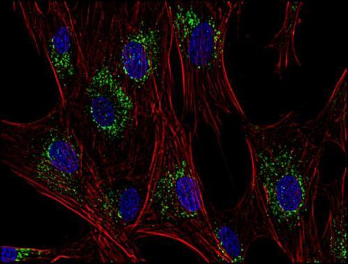
- Main image
- Experimental details
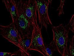
- Immunofluorescence staining of CD63 in human HeLa cell line using anti-CD63 (GTX28219; green). Actin cytoskeleton was labeled by phalloidin (red) and cell nuclei stained with DAPI (blue)
- Submitted by
- GeneTex (provider)
- Main image
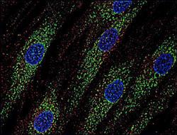
- Experimental details
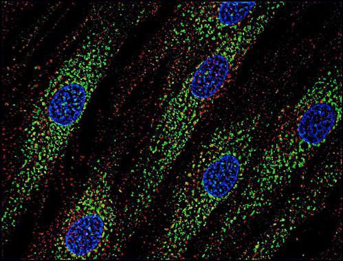
- Immunofluorescence staining of human skin fibroblasts with anti-CD63 (GTX28219; green) after co-incubation of living cells with human Transferrin - Dyomics 547 (red); cell nuclei stained with DAPI (blue)
Supportive validation
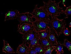
- Submitted by
- GeneTex (provider)
- Main image

- Experimental details
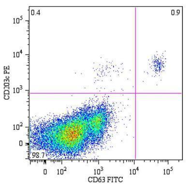
- Flow cytometry analysis of peripheral blood lymphocytes from a patient with allergy to bee venom after stimulation with bee venom, stained with anti-human CD63 [MEM-259] FITC (GTX18235)
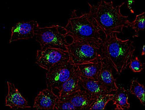
- Submitted by
- GeneTex (provider)
- Main image
- Experimental details
- Immunofluorescence staining of CD63 in human primary fibroblasts using anti-CD63 (GTX28219; green). Actin cytoskeleton was labeled by phalloidin (red) and cell nuclei stained with DAPI (blue)