 Explore
 Validate
 Learn
Learn Western blot
Antibody data
- Antibody Data
- Antigen structure
- References [1]
- Comments [0]
- Validations
- Western blot [2]
- Immunohistochemistry [4]
- Flow cytometry [3]
Submit
Validation data
Reference
Comment
Report error
- Product number
- TA802751 - Provider product page

- Provider
- OriGene
- Proper citation
- OriGene Cat#TA802751, RRID:AB_2626480
- Product name
- CD63 mouse monoclonal antibody, clone OTI2G6 (formerly 2G6)
- Antibody type
- Monoclonal
- Description
- CD63 mouse monoclonal antibody, clone OTI2G6 (formerly 2G6)
- Host
- Mouse
- Conjugate
- Unconjugated
- Epitope
- CD63
- Isotype
- IgG
- Antibody clone number
- OTI2G6
- Vial size
- 100 µl
- Concentration
- 1 mg/ml
Submitted references Mechanisms of the effectiveness of lipid nanoparticle formulations loaded with anti-tubercular drugs combinations toward overcoming drug bioavailability in tuberculosis.
Banerjee S, Roy S, Bhaumik KN, Pillai J
Journal of drug targeting 2020 Jan;28(1):55-69
Journal of drug targeting 2020 Jan;28(1):55-69
No comments: Submit comment
Supportive validation
- Submitted by
- OriGene (provider)
- Main image

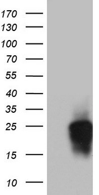
- Experimental details
- HEK293T cells were transfected with the pCMV6-ENTRY control (Left lane) or pCMV6-ENTRY CD63 (RC201733, Right lane) cDNA for 48 hrs and lysed. Equivalent amounts of cell lysates (5 ug per lane) were separated by SDS-PAGE and immunoblotted with anti-CD63.
- Validation comment
- WB
- Submitted by
- OriGene (provider)
- Main image

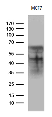
- Experimental details
- Western blot analysis of extracts (35ug) from MCF7 cell line by using anti-CD63 monoclonal antibody .(1:500)
- Validation comment
- WB
Supportive validation
- Submitted by
- OriGene (provider)
- Main image
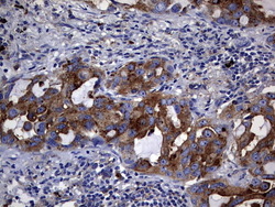
- Experimental details
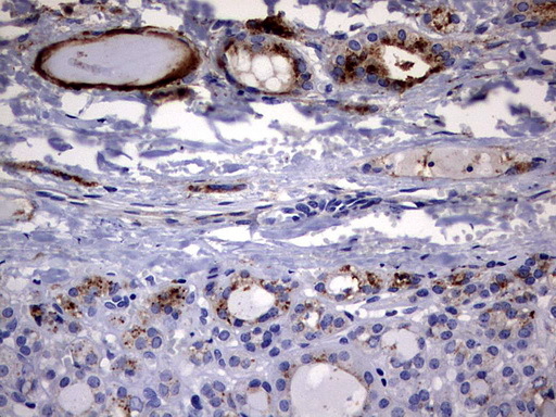
- Immunohistochemical staining of paraffin-embedded Carcinoma of Human lung tissue using anti-CD63 mouse monoclonal antibody. (Heat-induced epitope retrieval by 1 mM EDTA in 10mM Tris, pH9.0, 120C for 3min, TA802751)
- Validation comment
- IHC

- Submitted by
- OriGene (provider)
- Main image
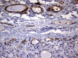
- Experimental details
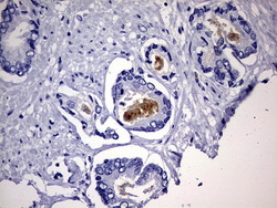
- Immunohistochemical staining of paraffin-embedded Carcinoma of Human thyroid tissue using anti-CD63 mouse monoclonal antibody. (Heat-induced epitope retrieval by 1 mM EDTA in 10mM Tris, pH9.0, 120C for 3min, TA802751)
- Validation comment
- IHC
- Submitted by
- OriGene (provider)
- Main image
- Experimental details
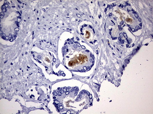
- Immunohistochemical staining of paraffin-embedded Human prostate tissue within the normal limits using anti-CD63 mouse monoclonal antibody. (Heat-induced epitope retrieval by 1 mM EDTA in 10mM Tris, pH9.0, 120C for 3min, TA802751)
- Validation comment
- IHC
- Submitted by
- OriGene (provider)
- Main image

- Experimental details
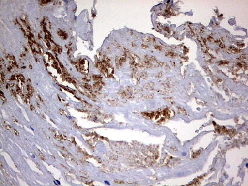
- Immunohistochemical staining of paraffin-embedded Carcinoma of Human bladder tissue using anti-CD63 mouse monoclonal antibody. (Heat-induced epitope retrieval by 1 mM EDTA in 10mM Tris, pH9.0, 120C for 3min, TA802751)
- Validation comment
- IHC
Supportive validation
- Submitted by
- OriGene (provider)
- Main image
- Experimental details
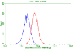
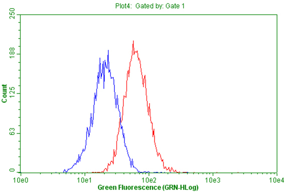
- Flow cytometric Analysis of living A549 cells,using anti-CD63 antibody(TA802751),(Red),compared to a nonspecific negative control antibody,(Blue).
- Validation comment
- FC
- Submitted by
- OriGene (provider)
- Main image

- Experimental details
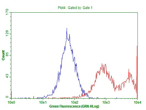
- HEK293T cells transfected with either RC201733 overexpress plasmid(Red) or empty vector control plasmid(Blue) were immunostained by anti-CD63 antibody(TA802751), and then analyzed by flow cytometry.(1:100)
- Validation comment
- FC
- Submitted by
- OriGene (provider)
- Main image
- Experimental details
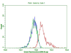
- Flow cytometric Analysis of MCF-7 cells, using anti-CD63 antibody(TA802751),(Red), compared to isotype control,(green), and negative control (PBS),(Blue)(1:100)
- Validation comment
- FC
