Explore
 Validate
 Learn
LearnPA5-17536
antibody from Invitrogen Antibodies
Targeting: CSNK1A1
CK1, CK1a, CK1alpha, CKIa, CKIalpha
 Western blot
Antibody data
- Antibody Data
- Antigen structure
- References [1]
- Comments [0]
- Validations
- Western blot [5]
- Other assay [1]
Submit
Validation data
Reference
Comment
Report error
- Product number
- PA5-17536 - Provider product page

- Provider
- Invitrogen Antibodies
- Product name
- CK1 alpha Polyclonal Antibody
- Antibody type
- Polyclonal
- Antigen
- Synthetic peptide
- Description
- It is not recommended to aliquot this antibody.
- Reactivity
- Human, Mouse, Rat
- Host
- Rabbit
- Isotype
- IgG
- Vial size
- 100 µL
- Concentration
- 46 µg/mL
- Storage
- -20°C
Submitted references Identification of modulators of autophagic flux in an image-based high content siRNA screen.
Hale CM, Cheng Q, Ortuno D, Huang M, Nojima D, Kassner PD, Wang S, Ollmann MM, Carlisle HJ
Autophagy 2016;12(4):713-26
Autophagy 2016;12(4):713-26
No comments: Submit comment
Supportive validation
- Submitted by
- Invitrogen Antibodies (provider)
- Main image

- Experimental details
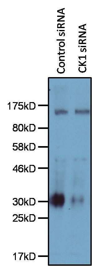
- Western blot analysis of CK1 was performed by loading 25 µg of whole cell lysates from HEK293T cells, treated with a control siRNA (left lane) or CK1 siRNA (right lane) for 48 hours, per well onto an SDS-PAGE gel. Proteins were transferred to a PVDF membrane and blocked with 5% milk in TBST for 30 minutes at room temperature. The membrane was probed with a CK1 polyclonal antibody (Product # PA5-17536) at a dilution of 1:500 overnight at 4C, washed in TBST, and probed with an HRP-conjugated anti-rabbit IgG secondary antibody at a dilution of 1:2000 for 1 hour at room temperature. Detection was performed using ECL Substrate (Product # 32106). Data courtesy of the Innovators Program.
- Submitted by
- Invitrogen Antibodies (provider)
- Main image

- Experimental details
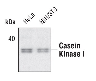
- Western blot analysis of CK1 in lysates from Hela and NIH/3T3 cell lines using CK1 polyclonal antibody (Product # PA5-17536).
- Submitted by
- Invitrogen Antibodies (provider)
- Main image
- Experimental details
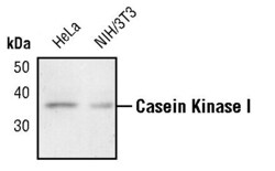
- Western blot analysis of CK1 in lysates from Hela and NIH/3T3 cell lines using CK1 polyclonal antibody (Product # PA5-17536).

- Submitted by
- Invitrogen Antibodies (provider)
- Main image
- Experimental details
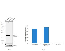
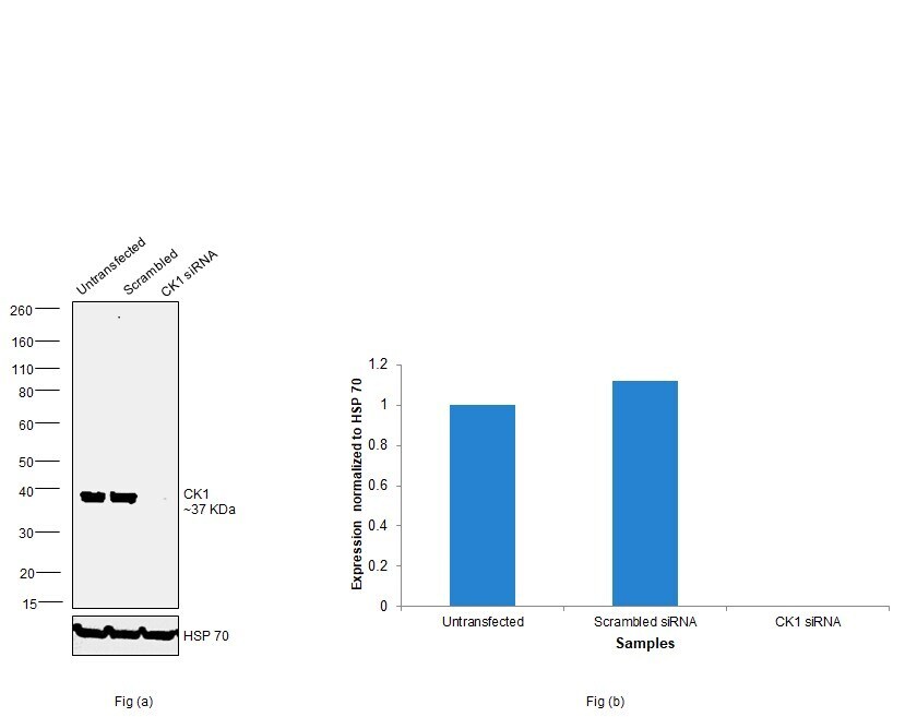
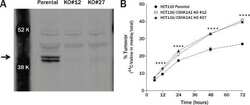
- Knockdown of CK1 was achieved by transfecting HEK-293 cells with CK1 specific siRNAs (Silencer® select Product # s3625). Western blot analysis (Fig. a) was performed using whole cell extracts from the CK1 knockdown cells (lane 3), non-specific scrambled siRNA transfected cells (lane 2) and untransfected cells (lane 1). The blots were probed with CK1 Polyclonal Antibody (Product # PA5-17536, 1:1000 dilution) and chemiluminescence with Goat anti-Rabbit IgG (H+L) Superclonal™ Recombinant Secondary Antibody, HRP (Product # A27036, 1:4000 dilution). Densitometric analysis of this western blot is shown in histogram (Fig. b). Decrease in signal upon siRNA mediated knock down confirms that antibody is specific to CK1
- Submitted by
- Invitrogen Antibodies (provider)
- Main image
- Experimental details
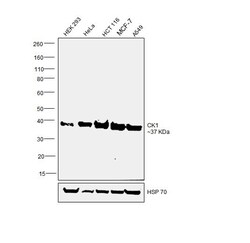
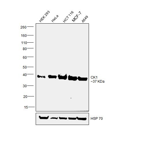
- Western blot was performed using anti-CK1 Polyclonal Antibody (Product # PA5-17536) and a 37 kDa band corresponding to CK1 was observed across cell lines tested. Whole cell extracts (30 µg lysate) of HEK 293 (Lane 1), HeLa (Lane 2), HCT 116 (Lane 3), MCF-7 (Lane 4) and A549 (Lane 5) were electrophoresed using NuPAGE™ 4-12% Bis-Tris Protein Gel (Product # NP0322BOX). Resolved proteins were then transferred onto a nitrocellulose membrane (Product # IB23001) by iBlot® 2 Dry Blotting System (Product # IB21001). The blot was probed with the primary antibody (1:1000 dilution) and detected by chemiluminescence with Goat anti-Rabbit IgG (H+L) Superclonal™ Recombinant Secondary Antibody, HRP (Product # A27036, 1:4000 dilution) using the iBright FL 1000 (Product # A32752). Chemiluminescent detection was performed using Novex® ECL Chemiluminescent Substrate Reagent Kit (Product # WP20005).
Supportive validation
- Submitted by
- Invitrogen Antibodies (provider)
- Main image
- Experimental details
- NULL