Explore
 Validate
 Learn
Learn Western blot
Antibody data
- Antibody Data
- Antigen structure
- References [0]
- Comments [0]
- Validations
- Western blot [3]
- Immunocytochemistry [1]
Submit
Validation data
Reference
Comment
Report error
- Product number
- PA1-4652 - Provider product page

- Provider
- Invitrogen Antibodies
- Product name
- DOPA Decarboxylase Polyclonal Antibody
- Antibody type
- Polyclonal
- Antigen
- Synthetic peptide
- Description
- This antibody is specific for the ~55 kDa DDC protein in Western blots of rat adrenal medulla.
- Reactivity
- Human, Rat, Bovine, Canine, Guinea Pig, Rabbit
- Host
- Rabbit
- Isotype
- IgG
- Vial size
- 100 µL
- Storage
- -20° C, Avoid Freeze/Thaw Cycles
No comments: Submit comment
Supportive validation
- Submitted by
- Invitrogen Antibodies (provider)
- Main image
- Experimental details
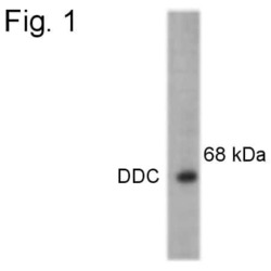
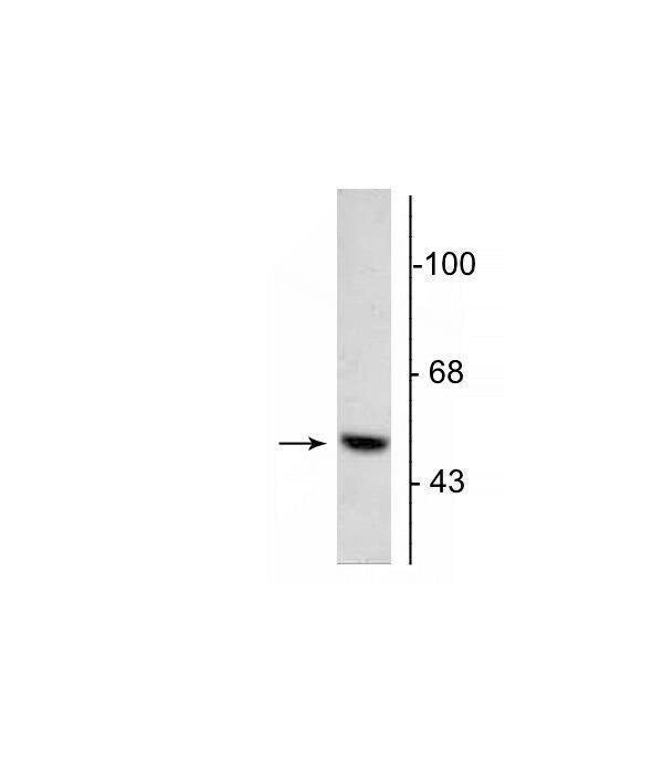
- Western blot of 5 µg of bovine adrenal medulla lysate showing specific immunolabeling of the ~55 kDa DOPA Decarboxylase protein.

- Submitted by
- Invitrogen Antibodies (provider)
- Main image

- Experimental details
- Western blot of DOPA Decarboxylase in bovine adrenal medulla lysate (5 µg). This shows specific immunolabeling of the ~55 kDa DOPA Decarboxylase polyclonal antibody (Product # PA1-4652).
- Submitted by
- Invitrogen Antibodies (provider)
- Main image
- Experimental details
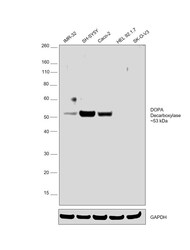
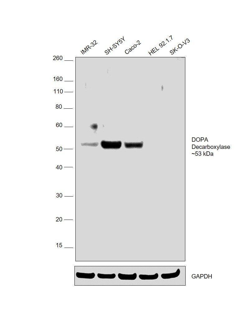
- Western blot was performed using Anti-DOPA Decarboxylase Polyclonal Antibody (Product # PA1-4652) and ~53 kDa band corresponding to DOPA Decarboxylase was observed in IMR-32, SH-SY5Y and Caco-2, but not in HEL.92.1.7 and SK-O-V3 which are reported to be negative for DOPA Decarboxylase expression. Whole cell lysate (40ug lysate) of IMR-32 (Lane 1), SH-SY5Y (Lane 2), Caco-2 (Lane 3), HEL 92.1.7 (Lane 4) and SK-O-V3 (Lane 5) were electrophoresed using Novex® NuPAGE® 4-12 % Bis-Tris gel (Product # NP0322BOX). Resolved proteins were then transferred onto a nitrocellulose membrane (Product # IB23001) by iBlot® 2 Dry Blotting System (Product # IB21001). The bot was probed with the primary antibody (1:1000 dilution) and detected by chemiluminescence with Goat anti-Rabbit IgG (H+L) Superclonal™ Recombinant Secondary Antibody, HRP (Product # A27036, 1:4000 dilution) using the iBright FL 1000 (Product # A32752). Chemiluminescent detection was performed using Novex® ECL Chemiluminescent Substrate Reagent Kit (Product # WP20005).
Supportive validation
- Submitted by
- Invitrogen Antibodies (provider)
- Main image
- Experimental details
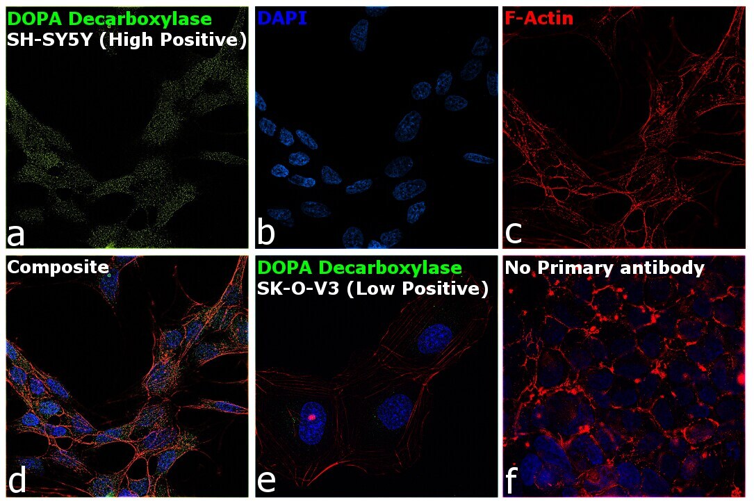
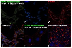
- Immunofluorescence analysis of DOPA Decarboxylase was performed using 70% confluent log phase SH-SY5Y and SK-O-V3 cells. The cells were fixed with 4% Paraformaldehyde for 10 minutes, permeabilized with 0.1% Triton™ X-100 for 10 minutes, and blocked with 2% BSA for 10 minutes at room temperature. The cells were labeled with DOPA Decarboxylase Polyclonal Antibody (Product # PA1-4652) at 1:100 dilution in 0.1% BSA, incubated at 4 degree celsius overnight and then labeled with Donkey anti-Rabbit IgG (H+L) Highly Cross-Adsorbed Secondary Antibody, Alexa Fluor Plus 488 (Product # A32790), (1:2000 dilution) for 45 minutes at room temperature (Panel a: Green). Nuclei (Panel b: Blue) were stained with SlowFade® Gold Antifade Mountant with DAPI (Product # S36938). F-actin (Panel c: Red) was stained with Rhodamine Phalloidin (Product # R415, 1:300). Panel d represents the merged image showing cytoplasmic and nuclear localization for DOPA Decarboxylase in SH-SY5Y cells. Panel e represents merged image of SK-O-V3, which is reported to be low expressing for the same. Panel f represents control cells with no primary antibody to assess background. The images were captured at 60X magnification.