 Explore
 Validate
 Learn
Learn Western blot
Antibody data
- Antibody Data
- Antigen structure
- References [0]
- Comments [0]
- Validations
- Western blot [8]
- Immunocytochemistry [2]
- Immunohistochemistry [2]
Submit
Validation data
Reference
Comment
Report error
- Product number
- PA5-34943 - Provider product page

- Provider
- Invitrogen Antibodies
- Product name
- GAP43 Polyclonal Antibody
- Antibody type
- Polyclonal
- Antigen
- Synthetic peptide
- Description
- Recommended positive controls: SK-N-SH, IMR32, SK-N-AS, mouse fetal brain, mouse hippocampus, rat hippocampus, mouse brain, rat brain.
- Concentration
- 1 mg/mL
No comments: Submit comment
Supportive validation
- Submitted by
- Invitrogen Antibodies (provider)
- Main image
- Experimental details
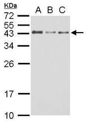
- Western blot analysis of GAP43 using A) 30 µg SK-N-SH whole cell lysate (B) 30 µg IMR32 whole cell lysate and C) 30 µg SK-N-AS whole cell lysate. Samples were loaded onto a 12% SDS-PAGE gel and probed with a GAP43 polyclonal antibody (Product # PA5-34943) at a dilution of 1:10000.

- Submitted by
- Invitrogen Antibodies (provider)
- Main image

- Experimental details
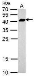
- Western blot analysis of GAP43 using A. 5 µg mouse brain lysate. Samples were loaded onto a 12% SDS-PAGE gel and probed with a GAP43 polyclonal antibody (Product # PA5-34943) at a dilution of 1:5000.

- Submitted by
- Invitrogen Antibodies (provider)
- Main image
- Experimental details
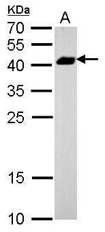
- Western blot analysis of GAP43 using A. 1 µg Rat brain lysate. Samples were loaded onto a 12% SDS-PAGE gel and probed with a GAP43 polyclonal antibody (Product # PA5-34943) at a dilution of 1:5000.
- Submitted by
- Invitrogen Antibodies (provider)
- Main image
- Experimental details
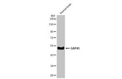
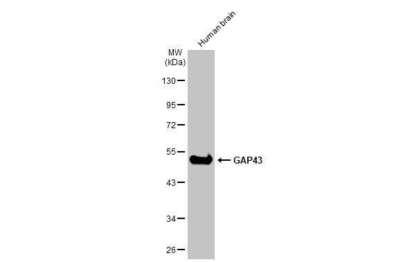
- Western Blot using GAP43 Polyclonal Antibody (Product # PA5-34943). Human tissue extract (30 µg) was separated by 10% SDS-PAGE, and the membrane was blotted with GAP43 Polyclonal Antibody (Product # PA5-34943) diluted at 1:30,000. The HRP-conjugated anti-rabbit IgG antibody was used to detect the primary antibody.
- Submitted by
- Invitrogen Antibodies (provider)
- Main image
- Experimental details
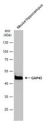
- Western blot analysis of GAP43 was performed by separating 50 µg of mouse tissue extract by 10% SDS-PAGE. Proteins were transferred to a membrane and probed with a GAP43 Polyclonal Antibody (Product # PA5-34943) at a dilution of 1:30000. The HRP-conjugated anti-rabbit IgG antibody was used to detect the primary antibody.

- Submitted by
- Invitrogen Antibodies (provider)
- Main image

- Experimental details
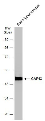
- Western blot analysis of GAP43 was performed by separating 50 µg of rat tissue extract by 10% SDS-PAGE. Proteins were transferred to a membrane and probed with a GAP43 Polyclonal Antibody (Product # PA5-34943) at a dilution of 1:30000. The HRP-conjugated anti-rabbit IgG antibody was used to detect the primary antibody.
- Submitted by
- Invitrogen Antibodies (provider)
- Main image

- Experimental details
- Western blot analysis of GAP43 was performed by separating 50 µg of various tissue extracts by 10% SDS-PAGE. Proteins were transferred to a membrane and probed with a GAP43 Polyclonal Antibody (Product # PA5-34943) at a dilution of 1:30000. The HRP-conjugated anti-rabbit IgG antibody was used to detect the primary antibody.

- Submitted by
- Invitrogen Antibodies (provider)
- Main image

- Experimental details
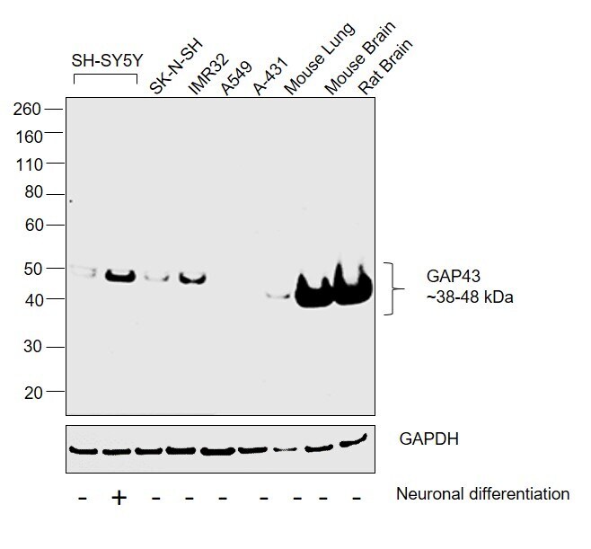
- Western blot was performed using Anti-GAP43 Polyclonal Antibody(Product # PA5-34943) and a 38-48 kDa band corresponding to GAP43 was observed across cell lines and tissues tested except in A549, A-431 and Mouse Lung which are reported to be low to negative for GAP43 expression. An increase in GAP43 expression was observed in SH-SY5Y on neuronal differentiation. Membrane enriched extracts (30 µg lysate) of SH-SY5Y (Lane 1), SH-SY5Y differentiated to neurons (Lane 2), SK-N-SH (Lane 3), IMR-32 (Lane 4), A549 (Lane 5), A-431 (Lane 6), Mouse Lung (Lane 7), Mouse Brain (Lane 8) and Rat Brain (Lane 9) were electrophoresed using NuPAGE™ 4-12% Bis-Tris Protein Gel (Product # NP0322BOX). Resolved proteins were then transferred onto a Nitrocellulose membrane (Product # IB23002) by iBlot® 2 Dry Blotting System (Product # IB21001). The blot was probed with the primary antibody (1:5000 dilution) and detected by chemiluminescence with Goat anti-Rabbit IgG (H+L) Superclonal™ Recombinant Secondary Antibody, HRP (Product # A27036,1:4000 dilution) using the iBright FL 1000 (Product # A32752). Chemiluminescent detection was performed using Novex® ECL Chemiluminescent Substrate Reagent Kit (Product # WP20005).
Supportive validation
- Submitted by
- Invitrogen Antibodies (provider)
- Main image

- Experimental details
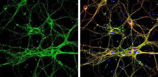
- Immunocytochemistry-Immunofluorescence analysis of GAP43 was performed in DIV9 rat E18 primary cortical neurons fixed in 4% paraformaldehyde at RT for 15 min. Green: GAP43 Polyclonal Antibody (Product # PA5-34943) diluted at 1:500. Red: beta Tubulin 3/ Tuj1, stained by beta Tubulin 3/ Tuj1 antibody. Blue: Fluoroshield with DAPI.
- Submitted by
- Invitrogen Antibodies (provider)
- Main image

- Experimental details
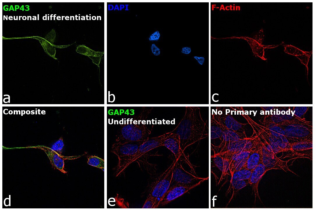
- Immunofluorescence analysis of GAP43 was performed using SH-SY5Y differentiated neuronal and undifferentiated cells. The cells were fixed with 4% paraformaldehyde for 10 minutes, permeabilized with 0.1% Triton™ X-100 for 15 minutes, and blocked with 2% BSA for 45 minutes at room temperature. The cells were labeled with GAP43 Polyclonal Antibody (Product # PA5-34943) at 1:200 dilution in 0.1% BSA, incubated at 4-degree Celsius overnight and then labeled with Donkey anti-Rabbit IgG (H+L) Highly Cross-Adsorbed Secondary Antibody, Alexa Fluor Plus 488 (Product # A32790), (1:2000 dilution), for 45 minutes at room temperature (Panel a: Green). Nuclei (Panel b: Blue) were stained with ProLong™ Diamond Antifade Mountant with DAPI (Product # P36962). F-actin (Panel c: Red) was stained with Rhodamine Phalloidin (Product # R415, 1:300). Panel d represents the merged image showing Plasma membrane and cytoplasm localization. Panel e represents undifferentiated SH-SY5Y cells with low to negative staining for GAP43. Panel f represents control cells with no primary antibody to assess the background. The images were captured at 60X magnification.
Supportive validation
- Submitted by
- Invitrogen Antibodies (provider)
- Main image

- Experimental details
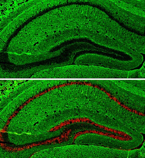
- Immunohistochemistry (Frozen) analysis of GAP43 was performed in frozen-sectioned adult mouse hippocampus tissue using GAP43 Polyclonal Antibody (Product # PA5-34943) at a dilution of 1:250 (Green). Red: NeuN, stained by NeuN antibody diluted at 1:500.
- Submitted by
- Invitrogen Antibodies (provider)
- Main image
- Experimental details
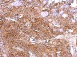
- GAP43 Polyclonal Antibody detects Gap43 protein at on Cal27 xenograft by immunohistochemical analysis. Sample: Paraffin-embedded Cal27 xenograft. GAP43 Polyclonal Antibody (Product # PA5-34943) dilution: 1:500. Antigen Retrieval: EDTA based buffer, pH 8.0, 15 min.
