 Explore
 Validate
 Learn
LearnPA1-25156
antibody from Invitrogen Antibodies
Targeting: CTNND1
CTNND, KIAA0384, p120, p120cas, p120ctn
 Western blot
Antibody data
- Antibody Data
- Antigen structure
- References [0]
- Comments [0]
- Validations
- Western blot [2]
- Immunocytochemistry [1]
Submit
Validation data
Reference
Comment
Report error
- Product number
- PA1-25156 - Provider product page

- Provider
- Invitrogen Antibodies
- Product name
- delta Catenin Polyclonal Antibody
- Antibody type
- Polyclonal
- Antigen
- Synthetic peptide
- Description
- Recommended positive controls: A431, ECV304.
No comments: Submit comment
Supportive validation
- Submitted by
- Invitrogen Antibodies (provider)
- Main image
- Experimental details
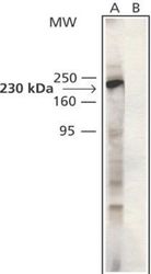
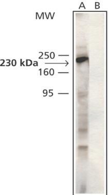
- Western blot analysis of delta Catenin in Cytosolic fraction of rat brain extract (Lanes A, C) and ECV304 whole cell extract (Lane B). Samples were probed with a delta Catenin polyclonal antibody (Product # PA1-25156). The antibody was developed with Anti-Rabbit IgG, Peroxidase and chemiluminescent substrate. A, B: Antibody dilution at 1:2000. C: Antibody dilution at 1:2000 + 20 µg/mL p120ctn immunizing peptide.
- Submitted by
- Invitrogen Antibodies (provider)
- Main image
- Experimental details
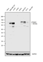
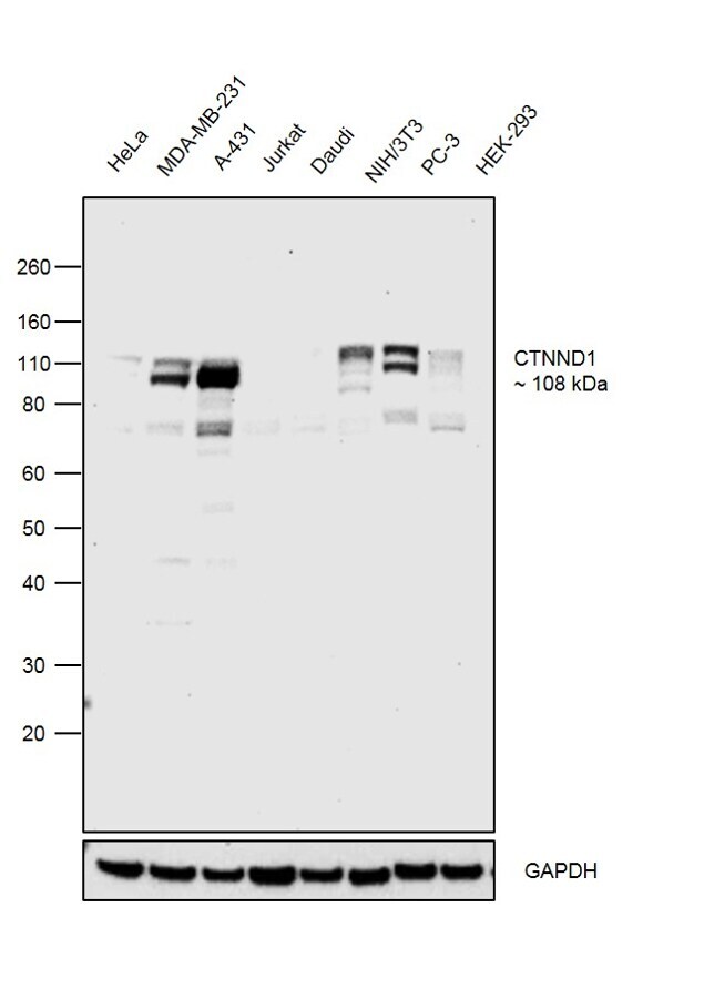
- Western blot was performed using Anti-delta Catenin Polyclonal Antibody (Product # PA1-25156) and a 108kDa band corresponding to CTNND1 was observed in all the tested cell models. Whole cell lysate (30ug lysate) of HeLa (Lane 1), MDA-MB-231 (Lane 2), A-431 (Lane 3), Jurkat (Lane 4), Daudi (Lane 5), NIH/3T3 (Lane 6), PC-3 (Lane 7) and HEK-293 (Lane 8) were electrophoresed using Novex® NuPAGE® 4-12 % Bis-Tris gel (Product # NP0322BOX). Resolved proteins were then transferred onto a nitrocellulose membrane (Product # IB23001) by iBlot® 2 Dry Blotting System (Product # IB21001). The blot was probed with the primary antibody (4ug/ml) and detected by chemiluminescence with Goat anti-Rabbit IgG (H+L), Superclonal™ Recombinant Secondary Antibody, HRP (Product # A27036, 1:4000 dilution) using the iBright FL 1000 (Product # A32752). Chemiluminescent detection was performed using SuperSignal™ West Dura Extended Duration Substrate (Product # 34076).
Supportive validation
- Submitted by
- Invitrogen Antibodies (provider)
- Main image
- Experimental details
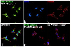
- Immunofluorescence analysis of delta Catenin was performed using 70% confluent log phase MDA-MB-231 cells. The cells were fixed with 4% paraformaldehyde for 10 minutes, permeabilized with 0.1% Triton™ X-100 for 15 minutes, and blocked with 2% BSA for 1 hour at room temperature. The cells were labeled with delta Catenin Rabbit Polyclonal Antibody (Product # PA1-25156) at 1:100 dilution in 0.1% BSA, incubated at 4 degree Celsius overnight and then labeled with Goat anti-Rabbit IgG (H+L) Superclonal™ Recombinant Secondary Antibody, Alexa Fluor® 488 conjugate (Product # A27034) at a dilution of 1:2000 for 45 minutes at room temperature (Panel a: green). Nuclei (Panel b: blue) were stained with SlowFade® Gold Antifade Mountant with DAPI (Product # S36938). F-actin (Panel c: red) was stained with Rhodamine Phalloidin (Product # R415, 1:300). Panel d represents the merged image showing plasma membrane and cytoplasmic localization. Panel e shows Daudi cells with no expression of delta Catenin. Panel f represents control cells with no primary antibody to assess background. The images were captured at 60X magnification.
