Explore
 Validate
 Learn
Learn Western blot
 Immunoprecipitation
Antibody data
- Antibody Data
- Antigen structure
- References [4]
- Comments [0]
- Validations
- Western blot [2]
- Immunocytochemistry [2]
Submit
Validation data
Reference
Comment
Report error
- Product number
- 33-7600 - Provider product page

- Provider
- Invitrogen Antibodies
- Product name
- p300 Monoclonal Antibody (NM-11)
- Antibody type
- Monoclonal
- Antigen
- Recombinant full-length protein
- Reactivity
- Human, Mouse
- Host
- Mouse
- Isotype
- IgG
- Antibody clone number
- NM-11
- Vial size
- 50 µg
- Concentration
- 0.5 mg/mL
- Storage
- -20°C
Submitted references Local BMP-SMAD1 signaling increases LIF receptor-dependent STAT3 responsiveness and primed-to-naive mouse pluripotent stem cell conversion frequency.
A SUMO-regulated activation function controls synergy of c-Myb through a repressor-activator switch leading to differential p300 recruitment.
The chromatin remodeling factor Mi-2alpha acts as a novel co-activator for human c-Myb.
Interferon-gamma interferes with transforming growth factor-beta signaling through direct interaction of YB-1 with Smad3.
Onishi K, Tonge PD, Nagy A, Zandstra PW
Stem cell reports 2014 Jul 8;3(1):156-68
Stem cell reports 2014 Jul 8;3(1):156-68
A SUMO-regulated activation function controls synergy of c-Myb through a repressor-activator switch leading to differential p300 recruitment.
Molvaersmyr AK, Saether T, Gilfillan S, Lorenzo PI, Kvaløy H, Matre V, Gabrielsen OS
Nucleic acids research 2010 Aug;38(15):4970-84
Nucleic acids research 2010 Aug;38(15):4970-84
The chromatin remodeling factor Mi-2alpha acts as a novel co-activator for human c-Myb.
Saether T, Berge T, Ledsaak M, Matre V, Alm-Kristiansen AH, Dahle O, Aubry F, Gabrielsen OS
The Journal of biological chemistry 2007 May 11;282(19):13994-4005
The Journal of biological chemistry 2007 May 11;282(19):13994-4005
Interferon-gamma interferes with transforming growth factor-beta signaling through direct interaction of YB-1 with Smad3.
Higashi K, Inagaki Y, Fujimori K, Nakao A, Kaneko H, Nakatsuka I
The Journal of biological chemistry 2003 Oct 31;278(44):43470-9
The Journal of biological chemistry 2003 Oct 31;278(44):43470-9
No comments: Submit comment
Supportive validation
- Submitted by
- Invitrogen Antibodies (provider)
- Main image
- Experimental details
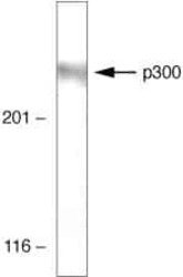
- Western blot analysis of HeLa cell lysates using p300 Monoclonal Antibody, Mouse/CBP (Product # 33-7600)

- Submitted by
- Invitrogen Antibodies (provider)
- Main image
- Experimental details
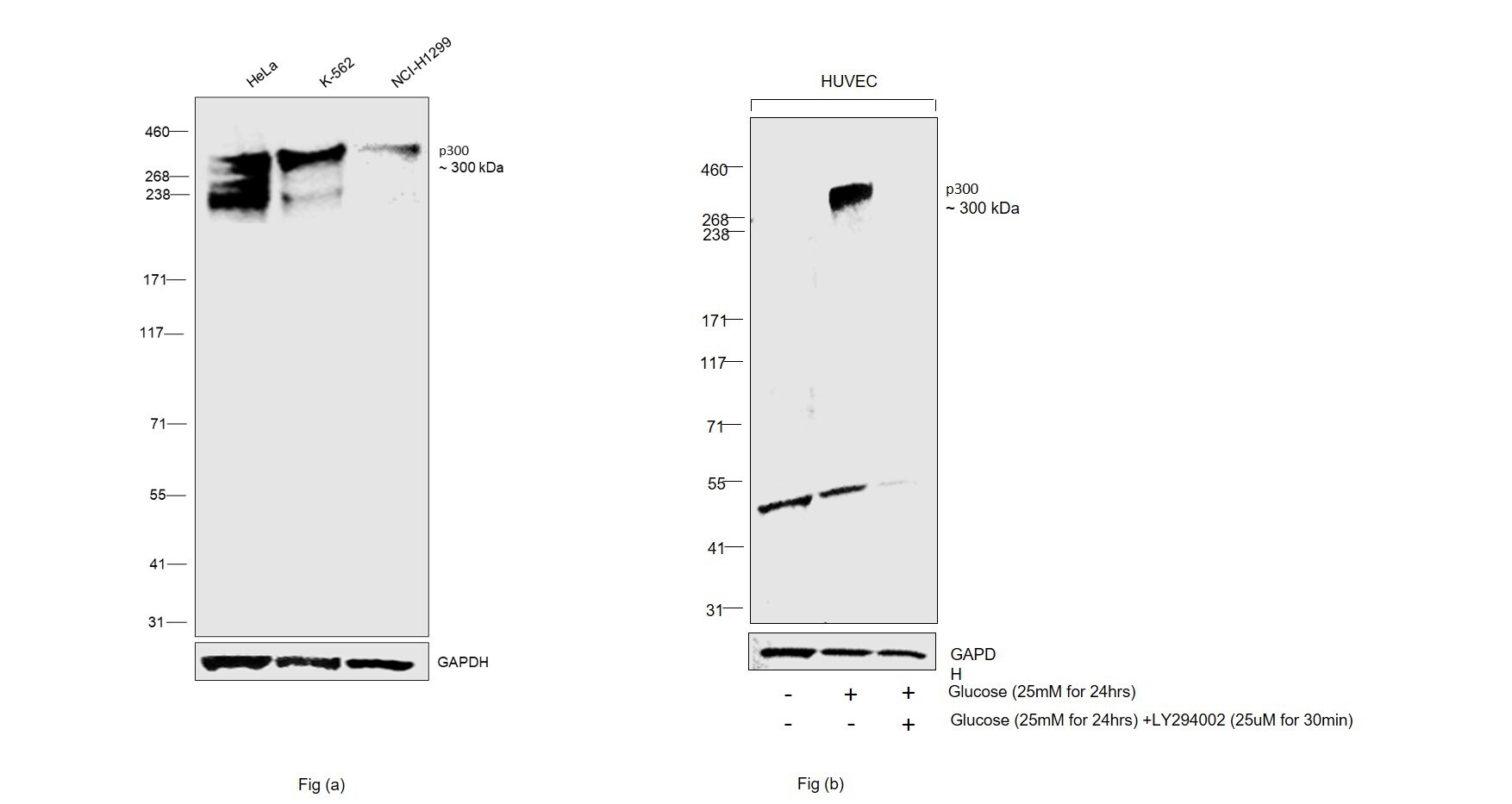
- Western blot was performed using Anti-p300 Monoclonal Antibody (NM-11) (Product # 33-7600) and a 300kDa band corresponding to p300 was observed across cell lines tested. Whole cell extracts (1%SDS) (40 µg lysate) of HeLa (Lane 1), K-562 (Lane 2), NCI-H1299 (Lane 3) as seen in Fig (a), HUVEC (Lane 1), HUVEC treated with Glucose (25mM for 24hrs) (Lane 2) and HUVEC treated with Glucose (25mM for 24hrs) and then treated with LY294002 (25uM for 30min) (Lane 3) as seen in Fig (b) were electrophoresed using NuPAGE™ 3-8% Tris-Acetate Protein Gel (Product # EA0378BOX). Resolved proteins were then transferred onto a nitrocellulose membrane (Product # IB23002) by iBlot® 2 Dry Blotting System (Product # IB21001). The blot was probed with the primary antibody (1:1000 dilution) and detected by chemiluminescence with Goat anti-Mouse IgG (H+L) Superclonal™ Recombinant Secondary Antibody, HRP (Product # A28177,1:20000 dilution) using the iBright™ FL1500 Imaging System (Product # A44115). Increase in signal was observed after stimulating HUVEC cells with Glucose and its expression was inhibited by using LY294002- an inhibitor, decrease in signal was observed upon treatment. Chemiluminescent detection was performed using SuperSignal™ West Pico PLUS Chemiluminescent Substrate (Product # 34580). (Ref :https://doi.org/10.2337/db06-0519).
Supportive validation
- Submitted by
- Invitrogen Antibodies (provider)
- Main image
- Experimental details
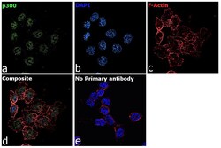
- Immunofluorescence analysis of p300 Monoclonal Antibody (NM-11) was performed using 70% confluent log phase HCT 116 cells. The cells were fixed with 4% paraformaldehyde for 10 minutes, permeabilized with 0.1% Triton™ X-100 for 15 minutes, and blocked with 2% BSA for 45 minutes at room temperature. The cells were labeled with p300 Monoclonal Antibody (NM-11) (Product # 33-7600) at 1:100 dilution in 0.1% BSA, incubated at 4 degree celsius overnight and then labeled with Donkey anti-Mouse IgG (H+L) Highly Cross-Adsorbed Secondary Antibody, Alexa Fluor Plus 488 (Product # A32766), (1:2000 dilution), for 45 minutes at room temperature (Panel a: Green). Nuclei (Panel b:Blue) were stained with ProLong™ Diamond Antifade Mountant with DAPI (Product # P36962). F-actin (Panel c: Red) was stained with Rhodamine Phalloidin (Product # R415, 1:300 dilution). Panel d represents the merged image showing nuclear as well as cytoplasmic localization. Panel e represents control cells with no primary antibody to assess background. The images were captured at 60X magnification.

- Submitted by
- Invitrogen Antibodies (provider)
- Main image

- Experimental details
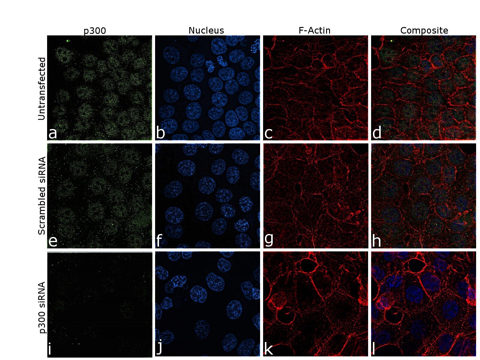
- Knockdown of p300 Monoclonal Antibody (NM-11) was achieved by transfecting HCT 116 cells with p300 specific siRNA (Silencer® select Product # S4696, S4697). Immunofluorescence analysis was performed on untransfected HCT 116 cells (panel a,d), transfected with non-specific scrambled siRNA (panels e,h) and transfected with p300 specific siRNA (panel i,l). Cells were fixed, permeabilized, and labelled with p300 Monoclonal Antibody (NM-11) (Product # 33-7600, 1:100 dilution) followed by Donkey anti-Mouse IgG (H+L) Highly Cross-Adsorbed Secondary Antibody, Alexa Fluor Plus 488 (Product # A32766), (1:2000 dilution). Nuclei (blue) were stained using ProLong™ Diamond Antifade Mountant with DAPI (Product # P36962), and Rhodamine Phalloidin (Product # R415, 1:300 dilution) was used for cytoskeletal F-actin (Red) staining. reduction in signal of specific signal was observed upon siRNA mediated knockdown (panel i,l) confirming specificity of the antibody to p300 (Green). The Images were captured at 60X magnification.