Explore
 Validate
 Learn
Learn Western blot
Antibody data
- Antibody Data
- Antigen structure
- References [0]
- Comments [0]
- Validations
- Western blot [4]
- Immunohistochemistry [1]
Submit
Validation data
Reference
Comment
Report error
- Product number
- PA5-111875 - Provider product page

- Provider
- Invitrogen Antibodies
- Product name
- SLC9A6 Polyclonal Antibody
- Antibody type
- Polyclonal
- Antigen
- Synthetic peptide
- Reactivity
- Human, Mouse, Rat
- Host
- Rabbit
- Isotype
- IgG
- Vial size
- 50 µL
- Concentration
- 0.8 mg/mL
- Storage
- -20°C
No comments: Submit comment
Supportive validation
- Submitted by
- Invitrogen Antibodies (provider)
- Main image

- Experimental details
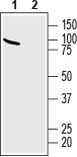
- Western Blot analysis of SLC9A6 was performed in human SH-SY5Y neuroblastoma cell line lysate. Lane 1: SLC9A6 Antibody (Product # PA5-111875) at a dilution of 1:200. Lane 2: SLC9A6 Antibody preincubated with the negative control antigen.
- Submitted by
- Invitrogen Antibodies (provider)
- Main image

- Experimental details
- Western Blot analysis of SLC9A6 was performed in human SH-SY5Y neuroblastoma cell line lysate. Lane 1: SLC9A6 Antibody (Product # PA5-111875) at a dilution of 1:200. Lane 2: SLC9A6 Antibody preincubated with the negative control antigen.
- Submitted by
- Invitrogen Antibodies (provider)
- Main image
- Experimental details
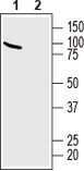
- Western Blot analysis of SLC9A6 was performed in mouse brain lysate. Lane 1: SLC9A6 Antibody (Product # PA5-111875) at a dilution of 1:200. Lane 2: SLC9A6 Antibody preincubated with the negative control antigen.

- Submitted by
- Invitrogen Antibodies (provider)
- Main image

- Experimental details
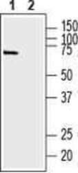
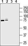
- Western Blot analysis of SLC9A6 was performed in rat brain membranes (lanes 1 and 3) and new born rat brain membranes (lanes 2 and 4). Lane 1,2: SLC9A6 Antibody (Product # PA5-111875) at a dilution of 1:200. Lane 3,4: SLC9A6 Antibody preincubated with the negative control antigen.
Supportive validation
- Submitted by
- Invitrogen Antibodies (provider)
- Main image

- Experimental details
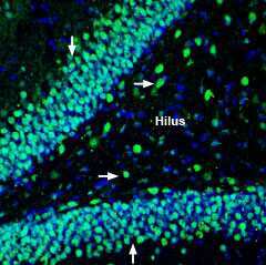
- Immunohistochemistry analysis of SLC9A6 in perfusion-fixed, frozen rat hippocampus tissue sections using SLC9A6 Antibody (Product # PA5-111875) at a dilution of 1:200, followed by goat-anti-rabbit-AlexaFluor-488. NHE-6 staining (green) in hippocampal dentate gyrus region, appears in neuronal soma outlines in the hilus (horizontal arrows) and the outer granule layer (vertical arrows). Cell nuclei were stained with DAPI (blue).