Explore
 Validate
 Learn
Learn Western blot
 ELISA
ELISAAntibody data
- Antibody Data
- Antigen structure
- References [0]
- Comments [0]
- Validations
- Western blot [2]
- Immunohistochemistry [1]
Submit
Validation data
Reference
Comment
Report error
- Product number
- NBP2-50217 - Provider product page

- Provider
- Novus Biologicals
- Product name
- Mouse Monoclonal CYP7B1 Antibody
- Antibody type
- Monoclonal
- Description
- Protein G purified.
- Reactivity
- Human
- Host
- Mouse
- Isotype
- IgG
- Vial size
- 0.1 ml
- Concentration
- 1 mg/ml
- Storage
- Store at 4C short term. Aliquot and store at -20C long term. Avoid freeze-thaw cycles.
No comments: Submit comment
Supportive validation
- Submitted by
- Novus Biologicals (provider)
- Main image

- Experimental details
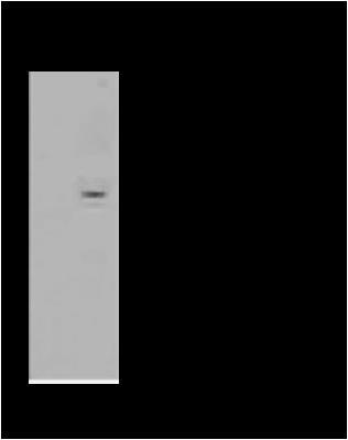
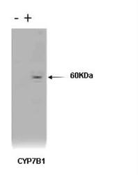
- Western Blot: CYP7B1 Antibody (M17-P3F2) [NBP2-50217] - Western blot was performed using anti-Cytochrome P450 7B1 [M17-P3F2]. The left hand lane contains control cell lysate while the right hand lane contains lysate prepared from cells over-expressing the protein. ~15 micrograms of protein were loaded per well. No bands were detected with the negative control lysate, while a band migrating at the expected molecular weight was observed in the over-expression lysate.
- Submitted by
- Novus Biologicals (provider)
- Main image
- Experimental details
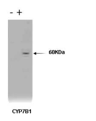
- Western Blot: CYP7B1 Antibody (M17-P3F2) [NBP2-50217] - Western blot was performed using anti-Cytochrome P450 7B1 [M17-P3F2]. The left hand lane contains control cell lysate while the right hand lane contains lysate prepared from cells over-expressing the protein. ~15 micrograms of protein were loaded per well. No bands were detected with the negative control lysate, while a band migrating at the expected molecular weight was observed in the over-expression lysate.
Supportive validation
- Submitted by
- Novus Biologicals (provider)
- Main image

- Experimental details
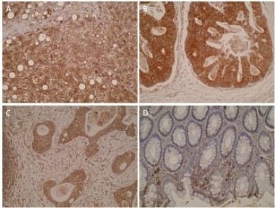
- Immunohistochemistry-Paraffin: CYP7B1 Antibody (M17-P3F2) [NBP2-50217] - Shows positive cytoplasmic immunostaining in normal liver (A) which is expected as CYP7B1 is involved in bile acid synthesis. Primary colorectal (B) and metastatic colorectal tumors (C) showed stronger staining compared to normal colon mucosa (D).