Explore
 Validate
 Learn
Learn Flow cytometry
Antibody data
- Antibody Data
- Antigen structure
- References [7]
- Comments [0]
- Validations
- Flow cytometry [1]
Submit
Validation data
Reference
Comment
Report error
- Product number
- 12-9117-41 - Provider product page

- Provider
- Invitrogen Antibodies
- Product name
- Anti-CD268 (BAFF Receptor) Monoclonal Antibody (8A7), PE, eBioscience™
- Antibody type
- Monoclonal
- Antigen
- Other
- Description
- Description: The 8A7 monoclonal antibody reacts with the human BAFF receptor, also known as B lymphocyte stimulator (BLyS protein) receptor, which is expressed on B cells. The ligand for this receptor, BAFF (B cell-activating factor of the TNF family) is a B cell survival factor and regulates CD21/35 and CD23 expression. Interaction of this ligand with its receptor causes elevated CD21/35 and CD23 expression, whereas receptor blockade has been shown to down-modulate expression. Applications Reported: The 8A7 antibody has been reported for use in flow cytometric analysis. Applications Tested: This 8A7 antibody has been pre-titrated and tested by flow cytometric analysis of normal human peripheral blood cells. This can be used at 5 µL (0.00375 µg) per test. A test is defined as the amount (µg) of antibody that will stain a cell sample in a final volume of 100 µL. Cell number should be determined empirically but can range from 10^5 to 10^8 cells/test. Excitation: 488-561 nm; Emission: 578 nm; Laser: Blue Laser, Green Laser, Yellow-Green Laser. Filtration: 0.2 µm post-manufacturing filtered.
- Reactivity
- Human
- Host
- Mouse
- Conjugate
- Yellow dye
- Isotype
- IgG
- Antibody clone number
- 8A7
- Vial size
- 25 Tests
- Concentration
- 5 µL/Test
- Storage
- 4° C, store in dark, DO NOT FREEZE!
Submitted references Type I Interferon Potentiates IgA Immunity to Respiratory Syncytial Virus Infection During Infancy.
IFN type I and II induce BAFF secretion from human decidual stromal cells.
BAFF promotes proliferation of human mesangial cells through interaction with BAFF-R.
The strength of the antibody response to the nematode Ascaris lumbricoides inversely correlates with levels of B-Cell Activating Factor (BAFF).
Chemokine receptor expression and functional effects of chemokines on B cells: implication in the pathogenesis of rheumatoid arthritis.
Altered B-cell homeostasis and excess BAFF in human chronic graft-versus-host disease.
Tonic B cell antigen receptor signals supply an NF-kappaB substrate for prosurvival BLyS signaling.
Hijano DR, Siefker DT, Shrestha B, Jaligama S, Vu LD, Tillman H, Finkelstein D, Saravia J, You D, Cormier SA
Scientific reports 2018 Jul 23;8(1):11034
Scientific reports 2018 Jul 23;8(1):11034
IFN type I and II induce BAFF secretion from human decidual stromal cells.
Lundell AC, Nordström I, Andersson K, Lundqvist C, Telemo E, Nava S, Kaipe H, Rudin A
Scientific reports 2017 Jan 6;7:39904
Scientific reports 2017 Jan 6;7:39904
BAFF promotes proliferation of human mesangial cells through interaction with BAFF-R.
Zheng N, Wang D, Ming H, Zhang H, Yu X
BMC nephrology 2015 May 15;16:72
BMC nephrology 2015 May 15;16:72
The strength of the antibody response to the nematode Ascaris lumbricoides inversely correlates with levels of B-Cell Activating Factor (BAFF).
Bornacelly A, Mercado D, Acevedo N, Caraballo L
BMC immunology 2014 Jun 7;15:22
BMC immunology 2014 Jun 7;15:22
Chemokine receptor expression and functional effects of chemokines on B cells: implication in the pathogenesis of rheumatoid arthritis.
Nanki T, Takada K, Komano Y, Morio T, Kanegane H, Nakajima A, Lipsky PE, Miyasaka N
Arthritis research & therapy 2009;11(5):R149
Arthritis research & therapy 2009;11(5):R149
Altered B-cell homeostasis and excess BAFF in human chronic graft-versus-host disease.
Sarantopoulos S, Stevenson KE, Kim HT, Cutler CS, Bhuiya NS, Schowalter M, Ho VT, Alyea EP, Koreth J, Blazar BR, Soiffer RJ, Antin JH, Ritz J
Blood 2009 Apr 16;113(16):3865-74
Blood 2009 Apr 16;113(16):3865-74
Tonic B cell antigen receptor signals supply an NF-kappaB substrate for prosurvival BLyS signaling.
Stadanlick JE, Kaileh M, Karnell FG, Scholz JL, Miller JP, Quinn WJ 3rd, Brezski RJ, Treml LS, Jordan KA, Monroe JG, Sen R, Cancro MP
Nature immunology 2008 Dec;9(12):1379-87
Nature immunology 2008 Dec;9(12):1379-87
No comments: Submit comment
Supportive validation
- Submitted by
- Invitrogen Antibodies (provider)
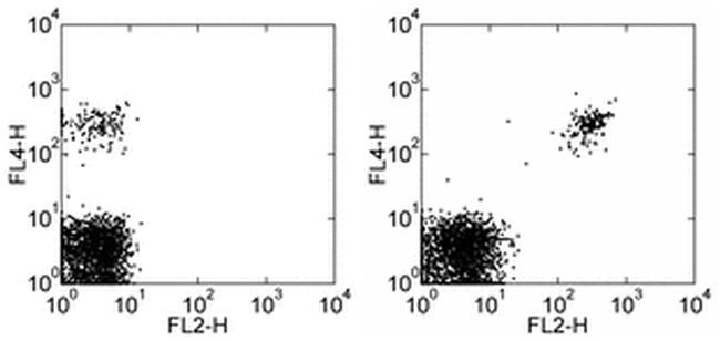
- Main image
- Experimental details
- Staining of normal human peripheral blood cells with Anti-Human CD19 APC (Product # 17-0199-42) and Mouse IgG2a kappa Isotype Control PE (Product # 12-4724-81) (left) or Anti-Human CD268 (BAFF Receptor) PE (right). Cells in the lymphocyte gate were used for analysis.
- Conjugate
- Yellow dye