 Explore
 Validate
 Learn
Learn Immunocytochemistry
Antibody data
- Antibody Data
- Antigen structure
- References [9]
- Comments [0]
- Validations
- Immunocytochemistry [2]
- Flow cytometry [1]
Submit
Validation data
Reference
Comment
Report error
- Product number
- MA5-13658 - Provider product page

- Provider
- Invitrogen Antibodies
- Product name
- ITGB1 Monoclonal Antibody (4B7R)
- Antibody type
- Monoclonal
- Antigen
- Other
- Description
- MA5-13658 targets CD29 in FACS, IF, and IHC (F) applications and shows reactivity with Human and Porcine samples.
- Antibody clone number
- 4B7R
- Concentration
- 0.2 mg/mL
Submitted references Potential localization of putative stem/progenitor cells in human bulbar conjunctival epithelium.
Cell adaptive response to extracellular matrix density is controlled by ICAP-1-dependent beta1-integrin affinity.
Telomerase-immortalized non-malignant human prostate epithelial cells retain the properties of multipotent stem cells.
Identification of putative stem cell markers, CD133 and CXCR4, in hTERT-immortalized primary nonmalignant and malignant tumor-derived human prostate epithelial cell lines and in prostate cancer specimens.
Gap junction protein connexin 43 serves as a negative marker for a stem cell-containing population of human limbal epithelial cells.
Re-arrangements of podosome structures are observed when Hck is activated in myeloid cells.
Partial enrichment of a population of human limbal epithelial cells with putative stem cell properties based on collagen type IV adhesiveness.
Characterization of putative stem cell phenotype in human limbal epithelia.
Basal cells of second trimester fetal breasts: immunohistochemical study of myoepithelial precursors.
Qi H, Zheng X, Yuan X, Pflugfelder SC, Li DQ
Journal of cellular physiology 2010 Oct;225(1):180-5
Journal of cellular physiology 2010 Oct;225(1):180-5
Cell adaptive response to extracellular matrix density is controlled by ICAP-1-dependent beta1-integrin affinity.
Millon-Frémillon A, Bouvard D, Grichine A, Manet-Dupé S, Block MR, Albiges-Rizo C
The Journal of cell biology 2008 Jan 28;180(2):427-41
The Journal of cell biology 2008 Jan 28;180(2):427-41
Telomerase-immortalized non-malignant human prostate epithelial cells retain the properties of multipotent stem cells.
Li H, Zhou J, Miki J, Furusato B, Gu Y, Srivastava S, McLeod DG, Vogel JC, Rhim JS
Experimental cell research 2008 Jan 1;314(1):92-102
Experimental cell research 2008 Jan 1;314(1):92-102
Identification of putative stem cell markers, CD133 and CXCR4, in hTERT-immortalized primary nonmalignant and malignant tumor-derived human prostate epithelial cell lines and in prostate cancer specimens.
Miki J, Furusato B, Li H, Gu Y, Takahashi H, Egawa S, Sesterhenn IA, McLeod DG, Srivastava S, Rhim JS
Cancer research 2007 Apr 1;67(7):3153-61
Cancer research 2007 Apr 1;67(7):3153-61
Gap junction protein connexin 43 serves as a negative marker for a stem cell-containing population of human limbal epithelial cells.
Chen Z, Evans WH, Pflugfelder SC, Li DQ
Stem cells (Dayton, Ohio) 2006 May;24(5):1265-73
Stem cells (Dayton, Ohio) 2006 May;24(5):1265-73
Re-arrangements of podosome structures are observed when Hck is activated in myeloid cells.
Poincloux R, Vincent C, Labrousse A, Castandet J, Rigo M, Cougoule C, Bordier C, Le Cabec V, Maridonneau-Parini I
European journal of cell biology 2006 Apr;85(3-4):327-32
European journal of cell biology 2006 Apr;85(3-4):327-32
Partial enrichment of a population of human limbal epithelial cells with putative stem cell properties based on collagen type IV adhesiveness.
Li DQ, Chen Z, Song XJ, de Paiva CS, Kim HS, Pflugfelder SC
Experimental eye research 2005 Apr;80(4):581-90
Experimental eye research 2005 Apr;80(4):581-90
Characterization of putative stem cell phenotype in human limbal epithelia.
Chen Z, de Paiva CS, Luo L, Kretzer FL, Pflugfelder SC, Li DQ
Stem cells (Dayton, Ohio) 2004;22(3):355-66
Stem cells (Dayton, Ohio) 2004;22(3):355-66
Basal cells of second trimester fetal breasts: immunohistochemical study of myoepithelial precursors.
Jolicoeur F, Gaboury LA, Oligny LL
Pediatric and developmental pathology : the official journal of the Society for Pediatric Pathology and the Paediatric Pathology Society 2003 Sep-Oct;6(5):398-413
Pediatric and developmental pathology : the official journal of the Society for Pediatric Pathology and the Paediatric Pathology Society 2003 Sep-Oct;6(5):398-413
No comments: Submit comment
Supportive validation
- Submitted by
- Invitrogen Antibodies (provider)
- Main image
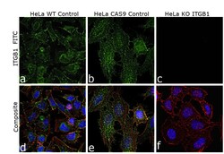
- Experimental details
- Knockout of ITGB1 was achieved by CRISPR-Cas9 genome editing using LentiArray™ Lentiviral sgRNA (Product # A32042, AssayID CRISPR626332_LV and CRISPR626355_LV) and LentiArray Cas9 Lentivirus (Product # A32064). Immunofluorescence analysis was performed on wild type HeLa cells (panel a,b) and HeLa ITGB1 KO cells (panel c,d). Cells were fixed, permeabilized, and labelled with ITGB1 Monoclonal Antibody (4B7R) (Product # MA5-13658, 1:100 dilution), followed by Donkey anti-Mouse IgG (H+L) Highly Cross-Adsorbed Secondary Antibody, Alexa Fluor Plus 488 (Product # A32766, 1:2,000). Nuclei (blue) were stained using ProLong™ Diamond Antifade Mountant with DAPI (Product # P36962), and Rhodamine Phalloidin (Product # R415, 1:300) was used for cytoskeletal F-actin (red) staining. Loss of signal (panel c,d) upon CRISPR mediated knockout (KO) confirms that antibody is specific to ITGB1 (green). The images were captured at 60X magnification.

- Submitted by
- Invitrogen Antibodies (provider)
- Main image
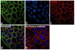
- Experimental details
- Immunofluorescence analysis of ITGB1 was performed using 70% confluent log phase A-431 cells. The cells were fixed with 4% paraformaldehyde for 10 minutes, permeabilized with 0.1% Triton™ X-100 for 15 minutes, and blocked with 2% BSA for 45 minutes at room temperature. The cells were labeled with ITGB1 Monoclonal Antibody (4B7R) (Product # MA5-13658) at 1:100 dilution in 0.1% BSA, incubated at 4 degree celsius overnight and then labeled with Donkey anti-Mouse IgG (H+L) Highly Cross-Adsorbed Secondary Antibody, Alexa Fluor Plus 488 (Product # A32766), (1:2000 dilution), for 45 minutes at room temperature (Panel a: Green). Nuclei (Panel b:Blue) were stained with ProLong™ Diamond Antifade Mountant with DAPI (Product # P36962). F-actin (Panel c: Red) was stained with Rhodamine Phalloidin (Product # R415, 1:300). Panel d represents the merged image showing Plasma membrane localization. Panel e represents control cells with no primary antibody to assess background. The images were captured at 60X magnification.

Supportive validation
- Submitted by
- Invitrogen Antibodies (provider)
- Main image
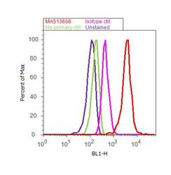
- Experimental details
- Flow cytometry analysis of Integrin beta-1 / CD29 was done on U-87 MG cells. Cells were fixed with 70% ethanol for 10 minutes, permeabilized with 0.25% Triton™ X-100 for 20 minutes, and blocked with 5% BSA for 30 minutes at room temperature. Cells were labeled with Integrin beta-1 / CD29 Mouse Monoclonal Antibody (MA513658, red histogram) or with mouse isotype control (pink histogram) at 3-5 ug/million cells in 2.5% BSA. After incubation at room temperature for 2 hours, the cells were labeled with Alexa Fluor® 488 Rabbit Anti-Mouse Secondary Antibody (A11059) at a dilution of 1:400 for 30 minutes at room temperature. The representative 10,000 cells were acquired and analyzed for each sample using an Attune® Acoustic Focusing Cytometer. The purple histogram represents unstained control cells and the green histogram represents no-primary-antibody control.
